Skip to content
Menu
Science News Learning
Incorporate STEM journalism in your classroom
Lesson plans
All guides
Search lesson plans
• Data Analysis
• Diversity in STEM
• Literacy Practices
• Research & Design
• Graph of the Week
Program info
Sign up
About
Testimonials
Community
Webinars
Journalism
Science News
Science News Explores
• Explainers
• Word of the Week
• Analyze This
• Technically Fiction
• Cool Jobs
• STEM Comics
Donate
SIGN IN
Science News Learning
Incorporate STEM journalism in your classroom
SIGN IN
Search
Open search
Close search
Incorporate STEM journalism in your classroom
Exercise type:
Activity
Topic:
Animals
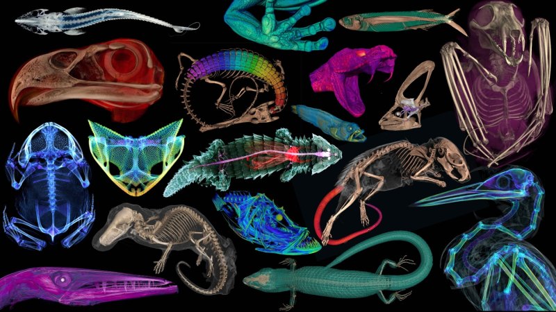
A peek inside
This exercise is a part of Educator Guide: Forest Neutrino Detectors and 3D Vertebrate Anatomy /
View Guide
Download Student Worksheet
With the completion of a yearslong project called openVertebrate, the insides of more than 13,000 museum specimens are seeing the...