Observing and analyzing an image

This exercise is a part of Educator Guide: Analyzing Images and an Exoplanet Collision Afterglow / View Guide
An image of the back of a rat's eye seen at 20 times magnification with immune cells seen in yellow and blood vessels seen in red.
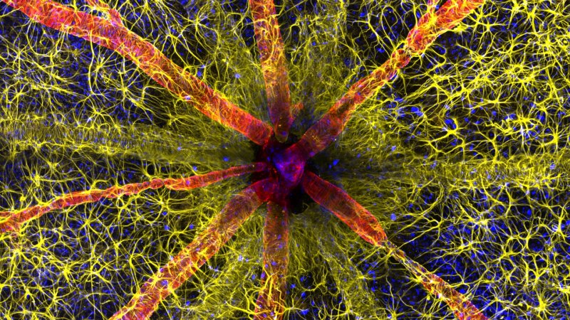
This image of the back of a rat’s eye, shown at 20 times magnification, showcases immune cells (yellow) and blood vessels (red) that populate the retina.Hassanain Qambari and Jayden Dickson

Directions for teachers

This short bellringer is designed for use with any image or visualization on the Science News or Science News Explores websites or print magazines.

Science News covers the annual Nikon Small World photo contest, which is a great resource for images that highlight microscopy’s role in exploring the natural world and its workings. Search the keyword “Nikon” on the Science News website to find results from different years. You can also search the Science News or Science News Explores homepage for current news images or search by a keyword for a topic-specific image.

Once you’ve chosen an image, remove its caption before sharing it with students. If you choose an online image for your class to analyze, post it to your online LMS and have students submit their responses. You can summarize student responses in a word cloud or in another type of data visualization. If you use a print image, ask students to shout out their answers and create a word cloud on a whiteboard. Or have students place sticky notes with their answers on the print image, clustering answers that are similar.

Share the caption with students before they answer questions 2-6 under “What it means.” You may need to add a sentence from the article that contains a summary of the research the image relates to. When you share this information with students, you can also give them the option to read the whole article if time allows.

Answers given below are based on the Science News article “The inside of a rat’s eye won the 2023 Nikon Small World photo contest” and refer to the winning photo.  

Directions for students

What I see

1. Look at the image your teacher has shared. What do you notice first? What do you notice as you spend more time looking at the image? Make at least two other observations about the image.

The first thing I notice is all the different colors in the image. The more I look at the image, the more details I see. There are red, tube-like structures that go from the center to the outside of the image. Yellow, wire-like structures swirl all around the space in the image and even seem to intersect the red tubes.

2. What initial questions do you have about the image?

Is this a magnification of a real structure or abstract art? Why is the inner part of the image darker than the rest? What do the colors mean?

What it means

1. What do you think the image is depicting? Explain your answer.

There appears to be a core to the image, with several things radiating from it. I think the image is of the center of something, like a cell.

Read the caption of the image before answering the following questions.

2. Write a brief description of what the image depicts. Were you correct in your guess? What questions, if any, do you still have about the image?

The image is of the back of a rat’s eye which has been magnified 20 times. The different colors depict different parts of the eye, including immune cells and blood vessels. I wasn’t correct that it was an image of a cell. I am wondering what the blue color represents.

3. How does the image help our understanding of its subject or a scientific concept?

The image shows that there are immune cells all over the imaged part of the retina. These immune cells are near the blood vessels. Researchers could be studying how the immune cells or blood vessels affect the health of the rat’s eye. The article describes that the image is a part of a research project focused on how diabetic retinopathy can change the structure and function of the retina. Similar images from animals with this disease may help scientists better understand the disease’s impact on retinas over time.

4. Would describing the image in words create the same impression? Explain.

Reading about the image wouldn’t be as impactful as seeing it. The magnified image with its artificial color allows the viewer to understand the distinct structures in the eye and their relative sizes and layout. It would be difficult to capture the information in words.  

5. What do you wonder about how the image was taken?

How was it possible to magnify the image so much? How did the photographer stage the rat to take the image? What was the strategy for adding color to the image, and how was that done?

6. What is a scientific concept or process that you’d like to photograph? How would an image help explain that concept or process?

Student answers will vary.