A new 3-D map illuminates the ‘little brain’ within the heart
Nerve cells in the organ are poorly understood
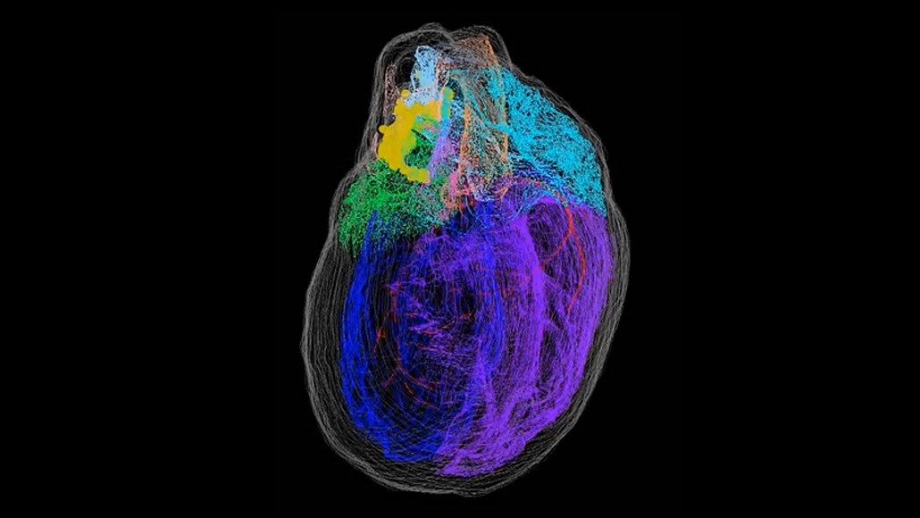
Nerve cells (yellow) that make up a heart’s “brain” cluster around the top of this reconstructed rat heart, near where blood vessels enter and exit the organ. Other colors show the contours of distinct heart areas, such as the left atrium (green), right atrium (teal), left ventricle (blue) and right ventricle (purple).
S. Achanta et al/iScience 2020