A deadly fungus gives ‘zombie’ ants a case of lockjaw
Closeups of infected ants’ jaw muscles may reveal clues to how the fungi take over
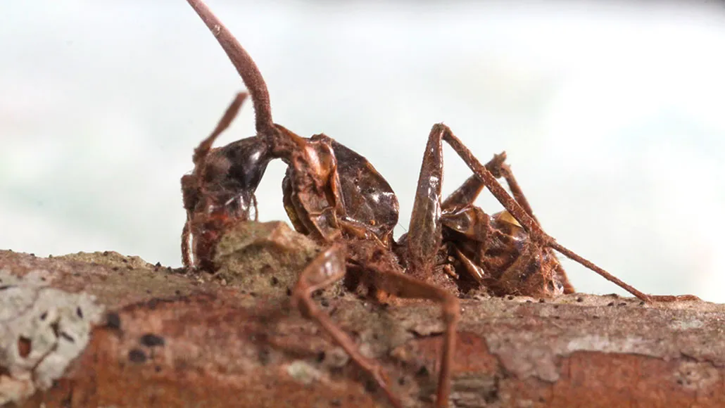
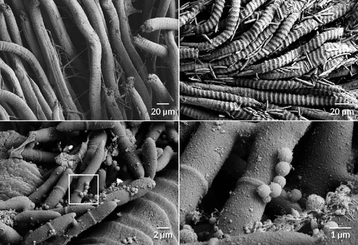
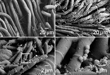
DEATH GRIP Once the “zombie ant fungus” invades an ant’s jaw muscles, the insect clings with clenched jaws to a twig until, and even after, its death.
Melissa Ishler; specimen collected by Kimberly Fleming.
Fungus-infected “zombie” ants are known to scale a plant, sink their jaws into a leaf or twig and wait to die while the Ophiocordyceps unilateralis fungi feast on the insects’ bodies. Eventually, a fungal stalk shoots out of the ant’s head and releases spores that rain down and infect more ants below.
The carpenter ants’ part in this nightmare may seem dictated by mind control, but the fungi don’t colonize the ants’ brains. Instead, the fungi take over ants’ jaws, forcing the muscles to contract into a death grip, researchers report July 17 in the Journal of Experimental Biology.
To unravel what exactly the fungus is doing to ants, scientists peered at infected ants’ stripy, striated jaw muscle fibers using scanning electron microscopy. “In infected muscles at the time of the death grip, … [the] lines appear really swollen,” says Colleen Mangold, a molecular biologist at Penn State University. The fungi wreck the muscle fibers but don’t seem to disturb the communication system that controls the muscles.
It’s still a mystery how the fungus initiates the death grip. But researchers may have found a clue: Tiny particles resembling clusters of grapes show up on infected muscle fibers.
Mangold and her colleagues think these particles may be extracellular vesicles, or packages of molecules, that are produced by either the invader or the host. If the orbs are vesicles, they may contain messages used by the fungi to take over ant bodies or play a role in the ants’ response, says Mangold.