Images probe artery-hardening plaques

Close look reveals which deposits pose heart attack risks

microscope image of plaques in a human artery

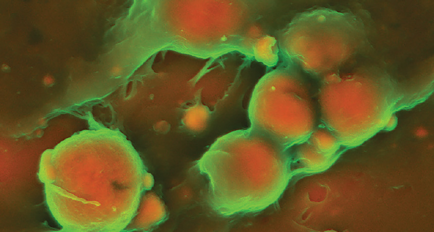
HAZARDOUS CLUMPS  The protein collagen (green) coats plaque (red) on the wall of a human artery. These small pockets of plaque are vulnerable to breaking away and possibly triggering a heart attack. 

J.D. Hutcheson et al/Nature Materials 2016

Collagen, best known as the key protein in skin, also protects against loose bits of plaque that can cause heart attacks. New images (below) of human arteries hardened by plaque illustrate the importance of collagen (green). The plaques (red and orange) in the left two micrographs are stable, while the plaques in the photos on the right are vulnerable to breaking away from the blood vessel wall.

Cells lining blood vessels secrete tiny spheres, called extracellular vesicles, filled with calcium and phosphate. The spheres fuse into calcified plaques (left images) under a protective collagen coating, Harvard Medical School’s Joshua Hutcheson and colleagues report January 11 in Nature Materials. The plaque poses little risk if the collagen layer isn’t disturbed. But when immune cells called macrophages invade the area to clear out infections or heal wounds, they can secrete chemicals that break down collagen and poke holes in the safety net. As a result, the vesicles clump into smaller, less stable plaques (right images).

Studying how arteries harden may lead to therapies that stabilize plaques and eventually dissolve them or prevent them from building up in the first place. 

Tina Hesman Saey is the senior staff writer and reports on molecular biology. She has a Ph.D. in molecular genetics from Washington University in St. Louis and a master’s degree in science journalism from Boston University.

More Stories from Science News on Life

From the Nature Index

Paid Content