A new nuclear imaging prototype detects tumors’ faint glow
Doctors could someday use Cerenkov light to detect cancer
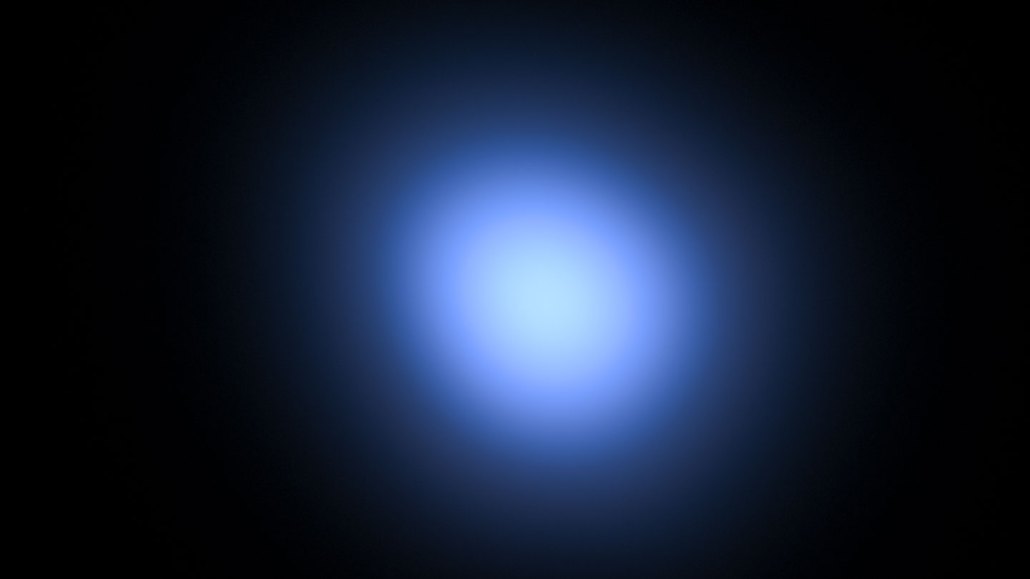
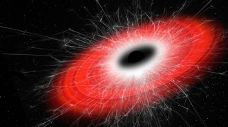
Cerenkov light, generated by high-speed particles traveling faster than light through a material, emits a blue glow. The light can be used to image a variety of cancers, new research shows.
Baac3nes/Moment/Getty Images Plus
A type of light commonly observed in astrophysics experiments and nuclear reactors can help detect cancer. In a clinical trial, a prototype of an imaging machine that relies on this usually bluish light, called Cerenkov radiation, successfully captured the presence and location of cancer patients’ tumors, researchers report April 11 in Nature Biomedical Engineering.
When compared with standard scans of the tumors, the Cerenkov light images were classified as “acceptable” or higher for 90 percent of patients, says Magdalena Skubal, a cancer researcher at Memorial Sloan Kettering Cancer Center in New York City.
Cerenkov radiation is generated by high-speed particles traveling faster than light through a material, such as bodily tissue (SN: 8/5/21). Nothing can travel faster than the speed of light in a vacuum, but light travels more slowly through a material, allowing particles to overtake it. In Cerenkov luminescence imaging, or CLI, particles released by radiotracers cause the target tissue to vibrate and relax in a way that emits light, which is then captured by a camera.
Between May 2018 and March 2020, in the largest clinical trial of its kind to date, 96 participants underwent both CLI and standard imaging, such as positron emission tomography/computed tomography, or PET/CT. Participants with a variety of diagnoses, including lymphoma, thyroid cancer and metastatic prostate cancer, received one of five radiotracers and were then imaged by the prototype — a camera in a light-proof enclosure.

Skubal and colleagues found that CLI detected all radiotracers, suggesting that the technology is more versatile than PET/CT scans, which work with only some radiotracers.
CLI images aren’t as precise as those from PET/CT scans. But CLI could be used as an initial diagnostic test or to assess the general size of a tumor undergoing treatment, says study coauthor Edwin Pratt, also of Memorial Sloan Kettering Cancer Center. “It would be a quick and easy way to see if there’s something off … [that warrants] further investigation,” Pratt says.
The findings strengthen the case for the technology as a promising low-cost alternative that could expand access to nuclear imaging in hospitals, says Antonello Spinelli, a preclinical imaging scientist at Experimental Imaging Centre in Milan, Italy, who was not involved in the research.
We summarize the week's scientific breakthroughs every Thursday.