X-ray vision
A new imaging technique could give scientists unprecedented views into cells and other objects at the nanoscale.
Scientists use X-rays to peer into a person’s body, and a new X-ray imaging technique does the same for individual cells.
“You should be able to image most macromolecular assemblies inside the cell”, such as proteins and DNA, says Pierre Thibault, a physicist at the Paul Scherrer Institute in Villigen, Switzerland who leads the team that reports the new technique in the July 18 Science.
Also, “it’s certainly a very important tool for nanotechnology,” Thibault says. The best light microscopes can’t distinguish features smaller than 200 nanometers. But images made with the new technique, called scanning X-ray diffraction microscopy, reveal features as small as 10 nanometers.“It’s an important development,” comments Jianwei Miao, a physicist at the University of California, Los Angeles who helped pioneer a related X-ray imaging technique. “The image is much better quality compared to previous ones.”
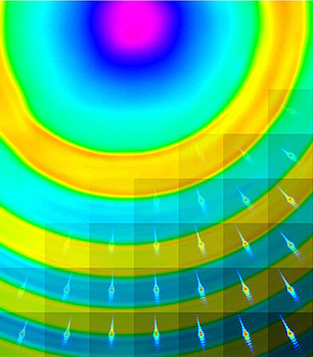
To make an image, Thibault and his colleagues scanned a specimen with an X-ray beam focused into a spot 300 nanometers across. For each point in the specimen, a high-speed photon detector recorded the X-rays that had passed through the specimen and spread out into a diffraction pattern — akin to the rainbow produced by a prism. Using the series of diffraction patterns, the scientists could mathematically reconstruct the image. “Our method should be easy to apply for 3-D imaging also,” Thibault adds.