A high-tech way to trace chemicals flowing through human nails might inspire better ways to fight fungus attacks.
Being tough as nails is a problem when treating fungal infections and other nail diseases. A nail’s tightly knit protein networks can block surface medications from reaching affected areas inside the nail. But an advanced imaging technology that can track chemicals’ movement through nails could help develop better treatments, researchers report online June 8 in Proceedings of the National Academy of Sciences.
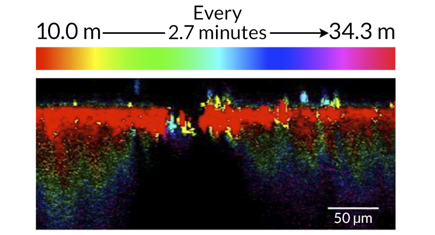
Improving drug formulas is a challenge, because it’s hard to measure how much of a drug a nail absorbs, says pharmaceutical chemist Richard Guy, of the University of Bath in England. Guy and colleagues, with collaborators at the University of Exeter in England, used an imaging technique called stimulated Raman scattering microscopy to make three-dimensional chemical maps of human nail clippings. The technique uses pulsing lasers to “vibrate” molecules. By tracking molecules’ unique vibrations, the scientists could follow a chemical’s motion through the nails.
“You’re looking at the real thing in real time,” says study coauthor and biophysicist Natalie Garrett of Exeter.
The researchers tested three liquids commonly found in pharmaceuticals — heavy water (D₂O), dimethyl sulfoxide, and propylene glycol. All three substances contained a heavy form of hydrogen, which allowed the scientists to clearly distinguish the chemicals’ vibrations from vibrations in the nail itself. Water penetrates fingernails quickly and deeply, the scientists report. In fact, water moved over 10 times faster than the other two chemicals and flowed twice as far.
Water’s small molecular size allows it to sneak through gaps in the nail’s protein network, Guy says. All three liquids moved more quickly across the nail as greater amounts were absorbed. Guy believes the chemicals loosened the nail’s protein network while sliding through.
The results will help inform drug design, Guy says. “We can now think about how we can formulate something … to keep the drug dissolved on the nail surface, so that it can be continuously provided to the nail and make its way across,” he says. He says his group is already thinking of the best drugs to test.
This imaging technique has never been used on nails before, though it has been used on skin, says biomedical engineer Ji-Xin Cheng of Purdue University in West Lafayette, Ind. Cheng believes stimulated Raman scattering microscopy has great research potential. In addition to studying drug delivery, he says, the technique can be used to observe how single cells operate.