DNA twists and turns into interacting sections that determine what a cell does and when
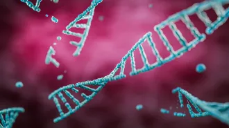
THE SHIFTING NUCLEAR TERRAIN Within the nucleus, as in this artist’s rendering, the genome coils around proteins and packs into a cell-specific architecture.
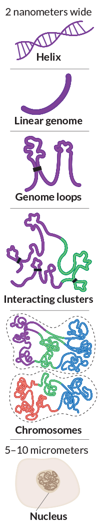
If you could unravel all the DNA in a single human cell and stretch it out, you’d have a molecular ribbon about 2 meters long and 2 nanometers across. Now imagine packing it all back into the cell’s nucleus, a container only 5 to 10 micrometers wide. That would be like taking a telephone cord that runs from Manhattan to San Francisco and cramming it into a two-story suburban house.
Fitting all that genetic material into a cramped space is step one. Just as important is how the material is organized. The cell’s complete catalog of DNA — its genome — must be configured in a specific three-dimensional shape to work properly. That 3-D organization of nuclear material — a configuration called the nucleome — helps control how and when genes are activated, defining the cell’s identity and its job in the body.
Researchers have long realized the importance of DNA’s precisely arranged structure. But only recently have new technologies made it possible to explore this architecture deeply. With simulations, indirect measurements and better imaging, scientists hope to reveal more about how the nucleome’s intricate folds regulate healthy cells. Better views will also help scientists understand the role that disrupted nucleomes play in aging and diseases, such as progeria and cancer.
PACKED TIGHT Within a cell nucleus, linear human DNA forms precise loops. Clusters of looped and coiled genetic material can interact with other nearby clusters. Such interactions occur within and between chromosomes, which are arranged in specific locations within the nucleus. Sources: S. Rao et al/Cell 2014; E. Lieberman-Aiden et al/Science 2009
“It is conceivable that every nuclear process has an element of structure in it,” says molecular geneticist Bing Ren of the University of California, San Diego School of Medicine. “It’s surprising, in fact, that we studied DNA for so long and yet we still have relatively little understanding of its 3-D architecture.”
Make that 4-D. Recent work shows that fully understanding the nucleome requires analysis of its rearrangements in space over time. A cell’s nucleome changes during the course of a single day as the cell responds to its environment.
Last year, the National Institutes of Health launched a five-year, 4-D Nucleome program, committing more than $120 million to identify better tools and techniques for mapping the complexities of the genome’s 4-D structure. Geneticists, molecular biologists, mathematicians, biophysicists and others are now on an ambitious quest to chart the ever-shifting nuclear terrain.
Location, location, location
The one-dimensional human genome is a biochemical instruction manual for building and operating a human being. Genetic instructions are written with four letters — A, T, C and G, abbreviations for the chemical subunits of DNA. The precise order of these letters encodes recipes for making the body’s proteins, as well as directions for how to use these biological building blocks.
But in the body, a genome is more than information written in DNA, says biophysicist William Greenleaf of Stanford University. It’s an interesting physical object.
In the nucleus of a human cell, the genetic material winds around protein spools, forming a net of DNA and protein called chromatin, which bundles into 46 parcels, the chromosomes. The chromatin arcs into thousands of loops, held in place by specialized proteins and the physical envelope of the nucleus. The genome manages to squeeze into its tight quarters without getting tangled like the earbuds cord in your pocket, says geneticist and computer scientist Erez Lieberman Aiden of Baylor College of Medicine in Houston.
For years, scientists have been trying to work out the rules for chromatin’s careful folding. In 2009, Aiden and his colleagues presented data in Science supporting a structure of dense, unknotted chromatin beads clustered into progressively larger clumps. A 2012 study in Proceedings of the National Academy of Sciences suggested that chromatin crumples into a range of different forms that vary on different chromosomes and over time.
While the nucleome may appear like tangled spaghetti, it’s more like a collection of structurally complex meatballs. Within the nucleus, chromosomes are arranged in specific locations. Each chromosome contains clusters of genetic material that get close enough, bundling together, to interact. These groupings can also engage with nearby clusters from other chromosomes.
Like the genetic text within it, the genome’s shape holds specific instructions. “The way it’s compacted forms this sort of physical memory of what the cell should be doing,” Greenleaf says.
Loops of DNA that aren’t needed by a particular cell are tucked away from the biological machinery that reads genetic blueprints, leaving only relevant genes accessible to produce proteins. Studies have shown that sections of the genome that are shoved toward the edges of a nucleus are often read less than centrally located DNA. Such specialized arrangements allow cells as diverse as brain cells, skin cells and immune cells to perform different jobs, even though each contains the same genome. “In different cell types, there are very large changes to the regions that are being used,” Greenleaf says.
Close contact
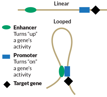
Source: J. Plank And A. Dean/Molecular Cell 2014; Credit: E. Otwell
Enhancers, genetic elements that increase gene activity, are often linearly far away from the genetic switches they control. When loops form, enhancers sidle up to their targets.
Sections of code that are separated by long stretches can wind up right next to each other, thanks to the looping. This positioning allows one part of the genome to interact with and control a gene that sits thousands of chemical letters away. For instance, enhancers — sections of DNA that increase a gene’s protein-producing activity — often seem to be far away from the genes that they control. But in three dimensions, enhancers cozy up to genetic switches that turn on a target gene. In a study published in Cell in 2014, Aiden and colleagues reported that roughly 30 percent of observed genome loops put an enhancer next to a far-off gene, complicating efforts to understand any gene’s regulation in isolation. “To understand such long-range interaction, we have to understand how DNA is folded,” Ren says.
Seeing is believing
Visualizing the structure of the nucleome has proved challenging. Microscopes can’t provide clear and complete images of nuclear architecture. But with modern DNA-reading technologies and the full text of the human genome, scientists are piecing together the genome’s three-dimensional shape.
A technique called Hi-C is one of the best options scientists have to scope out the contours of the 3-D genome. Developed by Aiden and his colleagues in 2009, Hi-C indirectly detects which sections of the genome are closest to one another in space inside a cell.
Scientists first chop the genome’s thread into small pieces within the nucleus, and then glue this genetic confetti back together with proteins. The proteins simply smash together any two DNA fragments that are sitting side-by-side, whether or not the fragments are close by in the actual sequence. The genome’s thread often gets patched back into its original order. But if two linearly distant swaths of genetic code were placed next to each other by a loop, these pieces may end up stuck together, a reflection of their three-dimensional proximity.
In Hi-C, scientists mark the newly paired genome chunks and then count how many times different genome sections are attached. When two distant sections of DNA are frequently observed stuck together, this suggests the presence of a loop.
In their 2014 study, Aiden and his colleagues used Hi-C to generate detailed spatial maps of the full genome in various human cells, including lung, breast and skin cells, as well as three types of cancer cells. The exploration uncovered thousands of genomic loops of varying size. Most are held in place by proteins called CTCF and cohesin. These proteins attach to a specific series of DNA letters that are arranged like bookends around the intervening genome. “The whole mechanism underlying this was sophisticated beyond anyone’s expectation,” Aiden says.
Story continues below infographic
Chop, chop
To identify sections of the genome that are spatially close to each other, the Hi-C method cuts DNA into pieces and reattaches neighboring pieces within the nucleus. Because bits of the genome that are nearby in 1-D are often nearby in 3-D, a partial Hi-C map (right) has intense values along the diagonal. Red dots at the corners of this map reveal distant areas of DNA on chromosome 13 that are joined by a loop.
Sources: S. Rao et al/Cell 2014; E. Lieberman-Aiden et al/Science 2009; credit: E. Otwell
But Hi-C imaging is just one tool for exploring the genome’s landscape. “The analogy would be to go around in life having earplugs and nose plugs and never touching anything,” he says. “You never want to rely on one sense; one sense can trick you.”
Mapping techniques that analyze proximity should be paired with imaging measurements to truly say something about the genome’s geometry, says Indika Rajapakse, a computational biologist at the University of Michigan in Ann Arbor. Rajapakse and his colleagues, for instance, try to paint a spatially accurate picture of the genome by pairing Hi-C with a technique nicknamed 3D-FISH, which labels DNA in three dimensions with glowing chemical tags.
While structural data may indicate which regions of the nucleome are interacting, even the sharpest map doesn’t say a thing about what such interactions mean for the cell, or how those interactions change. Scientists want to understand how the genome’s structure is connected to the genetic activities that build and control living beings — and this understanding requires a fourth dimension of analysis.
Time passages
A nucleome is constantly changing. “Time means that stimuli are happening,” Aiden says. A cell may change its activity as temperature changes, or as its human takes off for a run or goes to sleep.
To explore the link between a nucleome’s architecture and its shifting actions over time, Rajapakse and his colleagues generated numerical representations of the relationships between shape and function in the human genome over a 56-hour period. The team paired structural analysis with a technique that measures which DNA instructions are being read and followed at any given time.
Cyclical genes
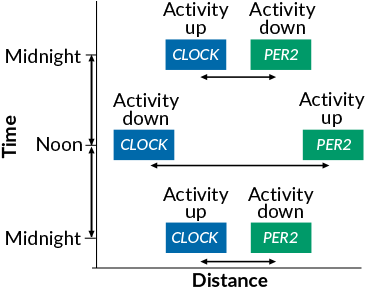
H. Chen et al/PNAS 2015, adapted by E. Otwell
In the nucleome, the circadian clock genes CLOCK and PER2 move toward and away from each other in a consistent 24-hour cycle, increasing and decreasing their activity as they go.
Nearly 2,000 genes shifted their shape, activity and position in relation to at least one other gene in the nucleome, the researchers reported in the Proceedings of the National Academy of Sciences in June. Two genes involved in regulating the body’s daily cycles (SN: 7/25/15, p. 24), CLOCK and PER2, perform a synchronized dance toward and away from each other in a dependable 24-hour pattern, increasing and decreasing their activity in opposition to one another. Even on Hi-C maps that reveal loops and connections of distant genome bits, the two genes are too far apart to show any physical contact.
The researchers say that existing structural analyses may miss important interactions between distant genes. Understanding the dynamic relationships between nuclear structure and genetic function, Rajapakse says, is the future of nucleome research. The scientists hope that mathematical analyses of the genome will identify important 4-D differences between various cell types and between healthy and diseased cells.
Shaping health
Scientists already know that disrupting the nucleome can cause disease. If the wrong sections of the genome end up next to each other, the controls intended for one gene may be applied to a different gene, with problematic results. In a study published in May in Cell, an international team of researchers produced limb malformations in mice by re-creating genetic alterations associated with hand and foot deformities in humans. Deleting or misplacing a chunk of genetic code can shift chromatin’s orientation, in this case resulting in fused, misshapen or extra digits, the team showed.
Altered nucleomes have also been linked to aging and aging disorders. Hutchinson-Gilford progeria syndrome, a fatal premature aging condition, results from mutations in the gene that encodes lamin A, a protein that normally supports the membrane surrounding the nucleus. In progeria, the nucleus becomes deformed and chromatin is damaged.
In June, a study in Sciencelinked disrupted nucleomes to a different premature aging condition, Werner syndrome (SN: 5/30/15, p. 13). Werner syndrome results when cells fail to produce working WRN protein, which, like lamin A, stabilizes a genome’s 3-D structure. As a result, young adults suffer symptoms such as osteoporosis, cataracts and hair loss.
Even healthy cells show a tight link between genome structure and aging: Older nucleomes accumulate genetic damage and appear to compact less tightly. In 2006, scientists found that naturally aging cells showed similar structural changes to cells ravaged by progeria.
Adjustments to the nucleome also play a role in cancer. In fact, in 1914 German biologist Theodor Boveri made the connection between cancer and genetics while observing cancer cells’ misshapen chromatin. Rearranging DNA from one chromosome to another can lead to tumors. Some genetic swaps are seen with high frequency in certain cancers, reflecting the close proximity of specific stretches of the genome.
There are whole classes of diseases where people are going to have aberrations of where their loops are.
— Erez Lieberman Aiden
In 2009, researchers identified eight genes that were moved away from their normal nuclear positions in breast cancer cells. The genes were so predictably misplaced that the scientists suggested looking at a gene’s position in the nucleus as a potential indicator of cancer.
The nucleome’s role in health and aging has given researchers a new framework to study human disease, Ren says. The field has reached a stage where genetics researchers can use insight from the nucleome to understand and guide their own experiments, Aiden says. “It’s not just for people who are really passionate about these 3-D genomics questions.”
A better understanding of the nucleome will profoundly impact medicine, he predicts. “There are whole classes of diseases where people are going to have aberrations of where their loops are.”
Much more remains to be understood about how a genome’s shape directs its activity. Future maps might zero in on functionally interesting regions of the genome, Greenleaf says. But he cautions there is also a benefit to unbiased, general exploration. Focusing on one location in the nucleome might lead researchers to miss important structural information elsewhere, he says.
Rajapakse says a close relationship between data collection and quantitative modeling will be necessary to gain a complete understanding of the nucleome.
“Data will guide us to build mathematical models, and then we can make predictions and go to the lab,” he says. His next projects will explore how to use nuclear structure and function to reprogram cellular systems, changing a nucleome’s organization to turn one cell type into another, he says.
Once they have the nucleome’s organizational rules in hand, some scientists, including Aiden, hope to engineer genomic loops in the lab. Because loops tightly control a gene’s activities, engineered loops could be used in gene therapies as a biological on/off switch for inserted genes, Aiden says.
High-quality maps of the nucleome are poised to uncover rich biological truths, Aiden says. He views the task ahead as similar to early astronomy. For Galileo to discover that Jupiter had moons, or that the Milky Way was a galaxy packed full of stars, he simply had to point his telescope in the right direction.
“I feel like in a small way, 3-D genome maps are eloquent in that way,” Aiden says. “Once you have that resolution…you can just read biology.”
This article appears in the September 5, 2015, issue of Science News with the headline, “The Shifting Nuclear Terrain: The human genome’s architecture is a moving target.”