Visualization offers view of a nerve cell’s dispatch center

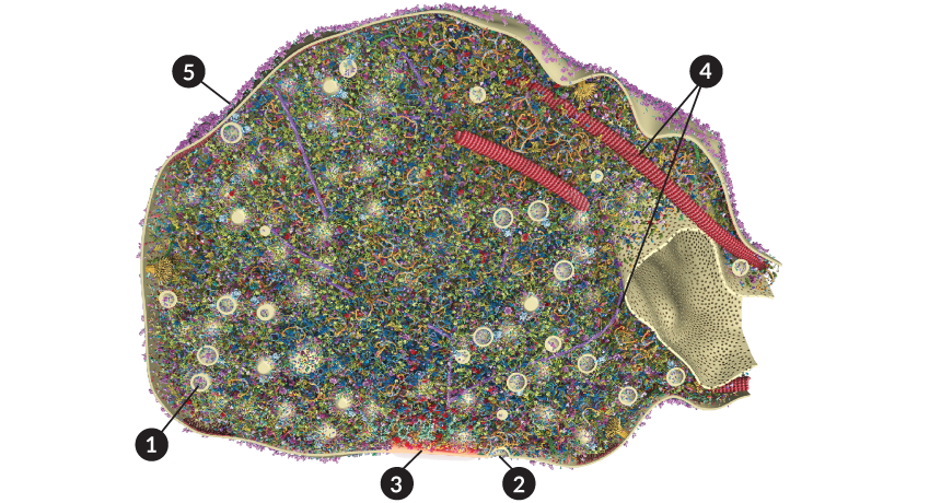
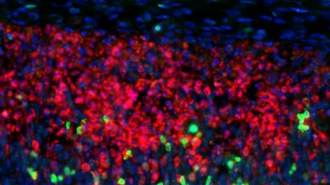
A 3-D visualization of the structure at the tip of a rat nerve cell's axon, called the synaptic bouton, reveals the machinery that allows neurons to send messages.

All: B.G. Wilhelm et al/Science 2014

See numbered detailsView the video

Brains run on constant streams of chatter. The roughly 85 billion nerve cells in the human brain converse by sending messages via molecules called neurotransmitters. These chemical conversations allow the brain to think, remember and feel, but the details of how those messages move remain mysterious.

To get a closer look, researchers led by Silvio Rizzoli of the University of Göttingen Medical Center in Germany created a 3-D visualization of the cellular machinery that allows a rat nerve cell to send a message. The new model, published in the May 30 Science, reveals the roles of proteins in the synaptic bouton, a structure at the tip of a nerve cell’s message-sending axon.

The model provides a clearer view of how nerve cells communicate. The team found, for example, that the synaptic bouton has an abundance of proteins that help send vesicles packed with neurotransmitters to the surface of the cell, but proteins that help retrieve the used vesicles are scarce. Rizzoli and his colleagues plan on building an even more ambitious model: “We want to go whole hog and do the whole neuron,” he says.

A closer look at the brain’s chatter

  • 1. A synaptic vesicle, about 42 nanometers wide, is loaded down with chemicals that serve as signals between nerve cells.
  • 2. After fusing with the bouton’s outer membrane and releasing its contents, a vesicle gets recycled back into the bouton.
  • 3. Vesicle melding is concentrated in the active zone (orange), a place where specialized proteins congregate to help the vesicles release their payloads.
  • 4. Microtubules (red) and actin filaments (purple) help give the synaptic bouton structure and move cargo around inside the neuron.
  • 5. The Alzheimer’s–related APP protein (purple) decorates the outside of the bouton’s membrane, where the protein may play a role in nerve cell communication.

A 3-D visualization of the structure at the tip of a rat nerve cell’s axon, called the synaptic bouton, reveals the machinery that allows neurons to send messages. Images: Allen Institute for Brain, EnCor Biotechnology Inc.; National Institute on Aging/NIH; Video: Wilhelm et al/Science 2014; Reported by Laura Sanders; Produced and narrated by Ashley Yeager

Laura Sanders is the neuroscience writer. She holds a Ph.D. in molecular biology from the University of Southern California.

More Stories from Science News on Neuroscience