See 3-D models of animal anatomy from openVertebrate’s public collection
More than 13,000 museum specimens were CT scanned as part of a six-year-long project

With the completion of a yearslong project called openVertebrate, the insides of more than 13,000 museum specimens are seeing the light of day. Digital reconstructions of CT scans (some shown) show the anatomy of fluid-preserved vertebrates, as well as last meals, yet-to-be-born offspring and more.
openVertebrate
Frog entrails, lizard scales and mouse tails, oh my.
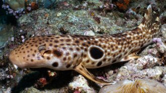
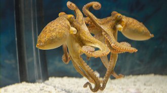
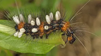
These creatures are among more than 13,000 museum specimens that had their innards CT scanned as part of a six-year mission to create 3-D digital reconstructions. The effort, called openVertebrate, or oVert, aims to make vertebrate specimens freely available online. Such specimens typically have been kept in storage until put on display for the public or pulled for examination by a specialist, researchers report March 6 in BioScience.
Online replicas not only make museum collections accessible to more folks but also give people a peek inside animals without the need for scalpels or other dissection equipment.
“The best part of that is the weird, wonderful things that you weren’t expecting to see that jump out,” says evolutionary biologist Edward Stanley of the Florida Museum of Natural History at the University of Florida in Gainesville. Those things include parasitic infections, last meals and new insights into animal anatomy.
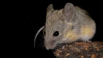
CT scans of pumpkin toadlets’ inner ears, for instance, revealed that the amphibians crash-land their hops due to misshapen ear tubes (SN: 6/15/22). And images that Stanley and colleagues took of spiny mice showed that the animals’ tails are covered in bony armor like an armadillo (SN: 5/24/23).
As part of oVert, Stanley and researchers across 25 institutions took CT scans of fluid-preserved specimens representing more than half of all known vertebrate genera, lighting up the skeletons of chameleons, frogs, bats, lizards, snakes, eagles and more. Some animals were soaked in iodine so that internal organs and muscles were visible.
A peek inside
As part of the openVertebrate project, researchers created an online repository of CT scans of more than 13,000 museum specimens. The goal was to make such collections more widely available to researchers without the need for travel or shipping precious specimens, as well as the public. Here’s a tiny sample of what those scans revealed.
-
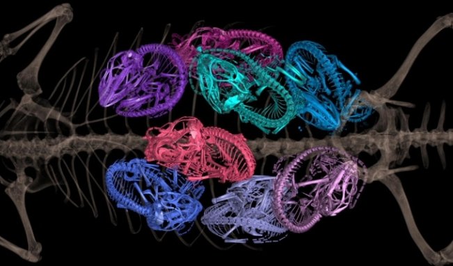
This spiny lizard (Sceloporus) was filled with eggs when it was preserved in fluid. CT scans lit up not just the mother’s bones, but also those of her eight offspring. openVertebrate -
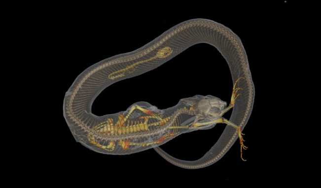
CT scans can provide a look into an animal’s recent diet without the need for dissection. This Eastern hognose snake (Heterodon platirhinos) specimen was not only swallowing a toad when it was preserved, it had recently chowed down on a salamander. openVertebrate -
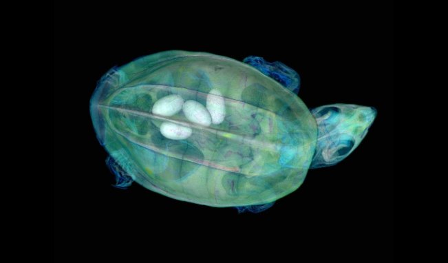
Turtles, including this Mexican musk turtle (Staurotypus triporcatus), required some creativity to scan. Their tough shells make it difficult to see the soft tissues beneath. Researchers had to directly inject iodine into turtles’ body cavities to produce CT scans of turtle soft tissue anatomy, including eggs. openVertebrate -
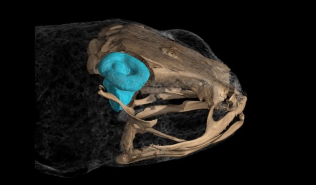
The inner ears of some Brachycephalus frogs are the smallest ever observed in vertebrates. But because ear tubes (shown in blue) take up proportionally more space within the frogs’ heads than in larger organisms, the amphibians are unable to balance while jumping. openVertebrate -

One of the most challenging animals to scan was an echidna (Tachyglossidae). These spiny anteaters presented a unique problem because their spikes made it difficult to fit the specimen back end first into the scanner’s tube, but its face was also too delicate to go face first. The team solved the problem by triple-bagging the organism in very tough plastic bags. openVertebrate
Each specimen was mounted inside a tube. The tube then rotated around a fixed X-ray scanner that captured a complete picture of the animal’s body. But few vertebrates are tube-shaped, so the team had to pack the cylinder with materials that could hold the specimen in place without interfering with the scan.
“It turns out bubble wrap, packing peanuts, plastic Coke bottles, that sort of thing, that’s the magic,” Stanley says.
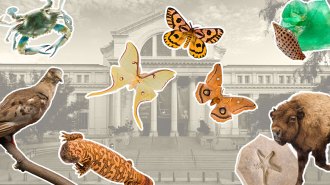
The technology could help digitize additional organisms tucked away in natural history collections including invertebrates and plants, the researchers say. Some scanners may even work for living vertebrates.