What a look at more than 3,000 kinds of cells in the human brain tells us
Analyzing single cells leads to new insights about how brains grow and operate
A deep look at the collections of cellular residents in the human brain offers hints about how it grows and works.
OsakaWayne Studios/Moment/Getty Images
A new look at the human brain is beginning to reveal the inner lives of its cellular residents.
The human brain holds a dizzying collection of diverse cells, and no two brains are the same, cellularly speaking. Those are the prevailing conclusions of an onslaught of 21 papers published online October 12 in Science, Science Advances and Science Translational Medicine.
The results just start to scratch the surface of understanding the mysteries of the brain. Still, they provide the most intimate look yet at the cells that build the brain, and offer clues about how the brain enables thoughts, actions and memories. The collection of data may also guide researchers in their hunt for the causes of brain disorders such as schizophrenia, Alzheimer’s disease and depression.
The new brain map is a result of a coordinated international research effort called the National Institutes of Health’s Brain Initiative Cell Census Network, or BICCN, which ramped up in 2017. Many of the studies in the collection are based on a powerful technology called single-cell genomics. The method reveals which genes are active inside of a single cell, information that provides clues about the cell’s identity and job.
As part of the BICCN, researchers examined all sorts of brains. One project detailed the cells in small pieces of live brain tissue taken from 75 people undergoing surgery for tumors or epilepsy, an approach that’s been used on smaller scales before (SN: 8/7/19). Another looked at samples taken from the brains of 17 deceased children. Still another looked at brain tissue from seven people, seven chimpanzees, four gorillas, three rhesus macaques and three marmosets.
The resolution provided by single-cell genomics revealed details about human brain cells in a way that previous methods couldn’t. “It’s remarkable how well it works,” says Ed Lein, a neuroscientist at the Allen Institute for Brain Science in Seattle and one of the lead researchers in the BICCN group. Collectively, the new studies describe over 3,000 cell types that reside in the human brain.
The main takeaway, Lein says, is that “the brain is really complex, from a cellular perspective.”
Amid that complexity, several key insights have already emerged, including hints about how human brains develop, how they vary among people, and how they differ from the brains of close primate relatives.
Growing brains
Some of the studies focused on very young brains. A study of the first two trimesters of brain growth, for instance, turned up previously unknown details about the identities of nerve cells in the thalamus, a kind of waystation for information coming into the brain. Many of those cells, called GABAergic neurons, are born elsewhere in the developing brain and migrate into the thalamus.
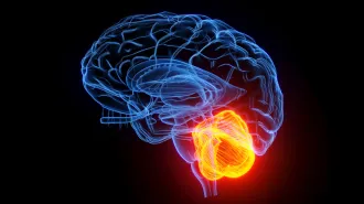
Other results show that the early years matter, a lot. Seth Ament, a neuroscientist at the University of Maryland School of Medicine in Baltimore, and his colleagues looked at brain cells in the cerebellum, a brain region at the back bottom of the brain. In children who died with inflammation in their brains, certain kinds of nerve cells there — Purkinje and Golgi neurons — had altered levels of active genes. The pattern, which held across eight brains, suggest that inflammation early in life could alter nerve cell development in certain spots.
“I’m amazed we saw something so consistent across the samples,” Ament says.
Unique brains
Some of the studies focused on variability between brain regions and between people.
One study looked at cells from about 100 spots taken from four adult brains. The researchers found, among many other things, that cells called astrocytes used their genes differently depending on where they reside. The finding hints that these cells, which are known to help nerve cells form connections and keep brain tissue healthy, may be specialized for their region.
Another study examined eight regions of the neocortex, the wrinkly outer area responsible for sophisticated thinking. Cells in those regions are somewhat standardized, sorting consistently into 24 categories, the scientists found. But the regions do have differences in the proportions of the cells. What that means for how these regions work is anybody’s guess.
Similarities also exist between people. Researchers found highly consistent patterns of cells when they compared brain cells from 75 people. But there was plenty of wiggle room, too. Microglia, immune cells in the brain that also sculpt nerve cell connections, were especially unique in the genes they use from person to person, for instance.
Primate brains
Some of the research compares human brains with primate relatives, including chimpanzees, gorillas, rhesus macaques and marmosets. By looking at cells in other primates’ brains, Lein says, “we finally get to ask the question about what makes humans unique.”
Overall, cells in the middle temporal gyrus, a part of the brain’s cortex, didn’t differ a lot between the primate brains. “It’s really remarkable that this complex cellular makeup is so conserved,” Lein says. “But you also have these changes.”
Compared with other primates, human brain cells use certain genes differently — in particular, genes related to how the cells form connections and communicate, researchers found. The analysis also turned up a few hundred genes that appear to behave in human-specific ways in brain cells. The researchers don’t yet know what those genes might be doing.
Imaging neuroanatomist Matthew Glasser cautions that it can be hard to tell exactly which brain areas are comparable among primates. Still, the results are “the first step in something really cool,” says Glasser, of Washington University School of Medicine in St. Louis, who was not involved in these studies.
Even better brain maps are coming
Overall, the progress represented by these and related results “is truly mind-blowing,” says cortical cartographer David Van Essen, who didn’t work on the new studies. “The community will certainly benefit from what’s coming out in this collection of papers.”
But, more importantly, it’s just a glimpse of what’s to come. “It’s not an end point in my view,” says Van Essen, also at Washington University. “It’s more a midpoint.”
Ament agrees. “These papers, as important as I think they are, are not the end,” he says. “It’s more the starting of it, and now we have a lot more work to do.”
The new brain maps will probably be revised, refined and added to, Lein says. Scientists are already working on the next iterations, which seek to combine zoomed-out views of brain networks and brain behavior with the ultrafine details provided by single-cell technology.
“Now that we have these techniques,” Glasser says, “we’re trying to combine them with brain imaging and systems neuroscience to actually try and figure out the puzzle.”