From the May 21, 1932, issue
GENES, ONCE HYPOTHETICAL, NOW SEEN AND PHOTOGRAPHED
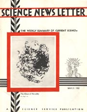
Genes, the ultimate units in heredity, have been seen and photographed. So declares Dr. John Belling, biologist on the staff of the Carnegie Institution of Washington.
Genes have hitherto been dealt with as hypothetical entities by biologists, because no one has ever actually seen them. They were like the atoms and electrons that make up matter: Physicists treat them as actually existing things, though it is impossible to given them visual demonstration. But now Dr. Belling believes that he has brought the genes out of their invisibility.
All living cells contain structures that presumably contain genes–the chromosomes within the nucleus. But to get clear-cut pictures of chromosomes, not all cells will do equally well. In the cells of some organisms chromosomes are too numerous or too small to work with conveniently; in others their outlines are not cleat-cut
Dr. Belling found lilies suitable for his purpose. By exceedingly fine and skillful microscopic technique, he got the contents of the pollen “mother-cells,” each only one four-hundredth of an inch in diameter, emptied out on glass slides. By suitable chemical treatment, he made the small divisions of the chromosomes, known as chromomeres, sharply visible. By further manipulation he was able to detect, within each chromomere, an exceedingly minute object, which he takes to be the gene itself. A typical cell of the type Dr. Belling has been working with contains about 4400 genes, arranged in 2200 pairs.
The picture on the cover of this weeks Science News Letter shows chromomere strings in a single cell of a lily.
X RAYS OF LIVER POSSIBLE THROUGH CHEMICAL INJECTION
Pioneer work in the use of a new method of diagnosing serious, often fatal diseases of the liver and spleen was reported to the American Medical Association meeting at New Orleans. The new method detected conditions that could not be determined by any other laboratory or clinical methods now in use, Dr. Wallace M. Yater, professor of medicine at Georgetown University School of Medicine, said in discussing the result of a study made by himself and his associate, Dr. Laurence S. Otell.
In using the new test, a small amount of a solution called thorium dioxide sol is injected into the veins every day for 3 days. On the fourth day X-ray pictures are taken of the liver and spleen. Ordinarily these important organs do not show up well on the X-ray plate, but after the thorium dioxide injections, the shape of both spleen and liver may be clearly seen. In this way physicians will be able to tell whether these organs are enlarged, whether there is fluid in the abdomen, whether such diseases as cancer, cirrhosis, or syphilis of the liver are present, and whether a large mass in the left side of the abdomen is an enlargement of the spleen or a tumor of some other organ.
SECRET OF PHOTOSYNTHESIS BAFFLING TO SCIENTISTS
Science has not yet solved the green leafs secret of storing up the energy of sunlight by converting carbon dioxide into carbohydrates, it appears from research by Prof. G. Mackinney of the University of Californias division of plant nutrition.
Vegetation has the ability of turning carbon dioxide, the gas exhaled by organisms and given off by fire, into carbohydrates, useful as starches, sugars and cellulose. Some 6 years ago Prof. E.C.C. Baly, professor of chemistry, University of Liverpool, reported the reduction of carbon dioxide to formaldehyde and carbohydrates in vitro, that is, in the test tube. Others worked on the same important problem with varying success. Prof. Mackinney has attempted to repeat the experiments but has been forced to conclude in his report to the American Chemical Society that “no procedure has yet been published whereby conditions for obtaining formaldehyde and carbohydrates in vitro can be duplicated.”
Photosynthesis, as this process is called, is fundamental to the existence of life on Earth through the utilization of sun energy.