A peek inside a turtle embryo wins the Nikon Small World photography contest

The annual competition highlights the power of microscopy to reveal hidden wonders

turtle embryo

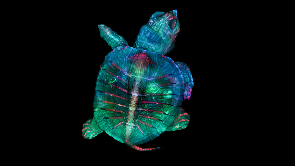
This image of a developing turtle embryo, stained to capture different tissues and shown at five times magnification, won the top award in the 2019 Nikon Small World photography contest.

Teresa Zgoda and Teresa Kugler/Nikon Small World

As scientists scour the natural world in search of truth, they often capture its beauty. 

Such was the case for microscopy technician Teresa Zgoda and micro-photographer Teresa Kugler. While helping with a class at the Marine Biological Laboratory in Woods Hole, Mass., the pair combined fluorescence microscopy and stereomicrography to create a vivid portrait of a developing turtle embryo. Because the embryo was more than 2.5 centimeters long, Zgoda, now at Brigham and Women’s Hospital in Boston, and Kugler, a recent graduate of the Rochester Institute of Technology in New York, couldn’t capture it all in such detail in one image. Instead, they stitched together hundreds of images focused on different locations and layers of the turtle to create a composite of the entire embryo.

The pair’s turtle image has won first place in the 2019 Nikon Small World photomicrography competition.

“Microscopy lets us zoom in on the smallest organisms and building blocks that comprise our world – giving us a profound appreciation for the small things in life that far too often go unnoticed,” Kugler said in an Oct. 21 news release announcing the results of the annual contest, now in its 45th year.

Here are some of our favorite entries among the other photo contest winners.

Stunning stentors

Dr. Igor Siwanowicz, a research scientist at the Howard Hughes Medical Institute’s Janelia Research Campus in Ashburn, Va., won second place for his vivid composite image of three single-celled microbes called stentors, sometimes known as “trumpet animalcules” for their horned shape. Their cilia, tiny hairs used for locomotion and feeding, stick out in contrast to the black background.

“I’m enamored with invertebrate morphology; usual evolutionary restraints don’t seem to apply within the realm of tiny animals, which is evident in the abundance and variety of often grotesque and utterly alien forms,” Siwanowicz wrote to the judging committee.

Siwanowicz says stentors are often difficult to see in their full, trumpet-like extended form because they rapidly change shape and contort their bodies. His solution to get the invertebrates to hold still: exposing them to magnesium ions. “Think a relaxing Epsom salt bath,” he says.

An image of three stentors, single-celled microorganisms common in freshwater lakes and streams, won second place in the competition. At 40 times magnification, each component of the cells is artificially stained.Igor Siwanowicz/HHMI

Hi, alligator

In Bhart-Anjan Bhullar’s paleontology and development lab at Yale University, graduate student Daniel Smith Paredes captured an American alligator embryo on day 20 of its development. 

Using different immunological stains, Smith Paredes was able to outline nerves (white) and their relationship to developing bone (yellow). He’s studying how a variety of vertebrate body forms arise during development. Like the winning entry, this image, which snagged third place, represents many thousands of individual pictures merged together.

American alligator embryo
Shown at 10 times magnification, this image of a developing American alligator embryo, which won third place in the contest, is stained to highlight developing bone (yellow) and nerves (white).Daniel Smith Paredes and Bhart-Anjan S. Bhullar/Yale University

Say freeze

Caleb Foster, a photographer in Jericho, Vt., snapped this photo of a snowflake on his back porch before it had time to melt. While snowflakes take a wonderful variety of forms, every snowflake has six sides because of how water molecules organize as they freeze. 

In the sky, ice crystals begin to form on a tiny speck of floating debris. The crystals then grow into their final configuration, influenced in part by the precise temperature and humidity around them. 

snowflake
All snowflakes have six sides, like this one pictured here at four times magnification.Caleb Foster/Caleb Foster Photography

Baby octopus

Carrie Albertin, a marine biologist at the Marine Biological Laboratory, studies how octopuses and other cephalopods build their strange bodies. She and Martyna Lukoševičiūtė, a scientist at the University of Oxford, submitted this image of a developing octopus embryo. Tiny tentacles, complete with suction cups, protrude from the main body of the creature.

Lukoševičiūtė told the judges that the biggest challenge in taking this photograph was getting the embryo to stay still. She used a specialized microscope slide with small grooves and walls to contain the embryo while she snapped its photo.

developing octopus
This image of a developing octopus was taken at five times magnification, and was artificially stained to visualize different tissues.Martyna Lukoševičiūtė/Univ. of Oxford and Carrie Albertin/MBL

Cherry cuprite

Emilio Carabajal Márquez from Madrid has studied cave insects for more than 25 years, but recently turned his eye toward microminerals found below Earth’s surface. Cuprite is a mineral composed of copper oxide, which gives the crystal a reddish color. This particular crystal may have been formed by lava vaporizing parts of an underground copper deposit.

cuprite
Cuprite, pictured here at 20 times magnification, is a crystallized mineral composed of copper oxide, which gives the cuprite a reddish color.Emilio Carabajal Márquez

Fly ovaries

Biologists Yujun Chen and Jocelyn McDonald of Kansas State University in Manhattan use fluorescent microscopy to help them understand how groups of cells get to where they need to go during development. In their image, the researchers captured a pair of fruit fly ovaries, stuffed with eggs at different stages of development. Muscle fibers are stained yellow while the nuclei of cells are green. Follicle cells, which support egg development, are outlined in purple.

“This image shows the… beauty of how different stages of egg chambers are packed and developed,” Chen told the judges.

fruit fly ovaries
A pair of fruit fly ovaries have been artificially stained and magnified 10 times to highlight the different stages of egg development.Yujun Chen and Jocelyn McDonald/Kansas State University

Pregnant plankton

Take a big gulp of lake water and there’s a pretty good chance you might swallow a daphnia. These tiny freshwater plankton use their featherlike mouthparts to feed on algae or other suspended organic material, and are an integral part of many freshwater ecosystems. Marek Miś, a freelance photographer in Suwalki, Poland, captured this pregnant daphnia. 

Daphnia can reproduce asexually, so there’s a chance that the developing daphnia (green) inside this female are clones of their mother.

pregnant Daphnia
This image of a pregnant female Daphnia, a microscopic invertebrate common in freshwater, was taken using polarized light to highlight the critter’s internal structures. The image is magnified four times.Marek Miś/Marek Miś Photography

Jonathan Lambert is a former staff writer for biological sciences, covering everything from the origin of species to microbial ecology. He has a master’s degree in evolutionary biology from Cornell University.

More Stories from Science News on Life

From the Nature Index

Paid Content