3-D printing builds bacterial metropolises

Technique may help researchers study antibiotic resistance

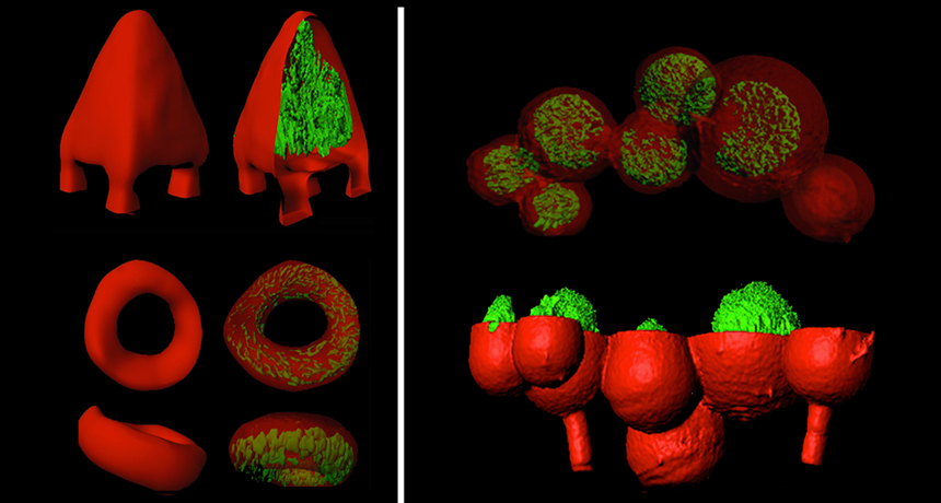
CAGED IN  Colonies of bacteria (green) nestle inside 3-D printed gelatin shells (red) in this computer-assisted 3-D microscopy image. Researchers can use 3-D printing to simulate social interactions between microorganisms.

J.L. CONNELL ET AL/ PROCEEDINGS OF THE NATIONAL ACADEMY OF SCIENCES 2013

Using a laser beam “printer” and globs of jelly as ink, scientists can now print tiny 3-D cities of bacteria in virtually any shape.

Bacteria can stick together to form slimy sheets called biofilms that yellow people’s teeth, line the lungs of cystic fibrosis patients and often resist antibiotics. Building 3-D models of bacterial communities could help explain how the microbes work together and evade drugs, says bioengineer Jason Shear of the University of Texas at Austin.

Shear and colleagues mixed bacteria with a drop of gelatin and a light-activated chemical glue. Then they hit the droplet with a laser to bind the gelatin together, forming a thin skin. Parts not touched by the laser washed away, leaving behind a hollowed-out space where the microbes could grow.

Researchers sealed bacteria inside gelatin boxes, doughnuts and pyramids, shapes that could mimic real biofilm structures. Shear’s team even nested one type of bacteria within a shell of another. When the team dosed the duo with an antibiotic, the exterior bacteria acted like a shield: It broke down the drug and protected the interior microbes from harm, the team reports October 7 in the Proceedings of the National Academy of Sciences.

Meghan Rosen is a staff writer who reports on the life sciences for Science News. She earned a Ph.D. in biochemistry and molecular biology with an emphasis in biotechnology from the University of California, Davis, and later graduated from the science communication program at UC Santa Cruz.

More Stories from Science News on Life

From the Nature Index

Paid Content