Elyse G.’s brain is fabulous. It’s also missing a big chunk
A new project explores interesting brains to better understand neural flexibility

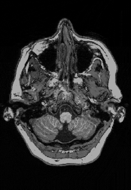
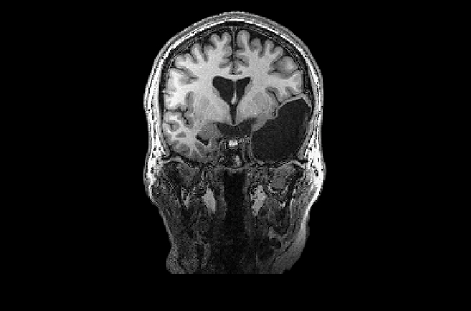
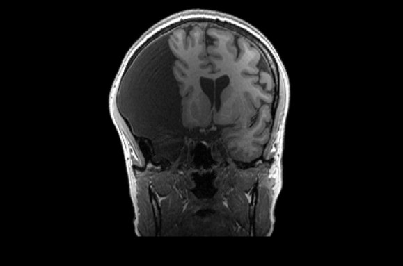
Elyse G. found out she lacked most of her left temporal lobe when she was 25 years old. The brain region is typically crucial for speech and language — but until her brain was scanned (three views shown), no one knew Elyse was missing hers.
COURTESY OF THE FEDORENKO LAB
EDITOR’S NOTE: Most of the Interesting Brains Project participants featured in this story are using shortened forms of their names and/or pseudonyms to protect their privacy.
You’d never guess that Elyse G. has a black hole in her brain.
Meet her on the street, and it’d be impossible to tell she’s lacking a chunk of neural tissue about the size of a small fist.
Looking at her brain scans is a different story. It’s as if someone has knocked over a bottle of ink. The darkness pools inside her skull near her left ear, a puddle of fuliginous black. Inside the splotch, there’s no white matter or gray matter, no blood vessels or tissue at all.
Elyse says you don’t have to be a neurosurgeon to spot what’s different about her brain: “There’s a big honking piece missing!”
Scientists can’t say exactly how it happened. It’s possible that sometime long ago, perhaps due to a stroke before or shortly after birth, a portion of Elyse’s brain died and then ultimately disappeared, leaving behind only liquid — brain tissue swapped for a fluid-filled void. Her sister has one too.
Elyse and her sister, Martha M., who are not using their full names to maintain their anonymity, look and act perfectly ordinary. But each lacks most of a temporal lobe, and each in a different hemisphere. Elyse is also missing part of her brain stem. The women are two of who knows how many people living their lives without brain structures generally thought to be crucial.
Martha, now age 59, didn’t know her brain was different until she was a teenager. Elyse, who will turn 61 this year, found out in graduate school. Two sisters. Two brains. Two black holes. When MIT cognitive neuroscientist Evelina Fedorenko’s team first learned about the duo, “we were all kind of blown away,” she says.
Elyse emailed Fedorenko her brain images in 2016, decades after the void was discovered. She had read an article about neuroscience research at MIT and was curious if scientists would be interested. “She said, ‘I’m missing my left temporal lobe. Do you want to study me?’ ” Fedorenko remembers.
The left temporal lobe is generally thought to be essential for speech and language, and Fedorenko, who trained as a language researcher, was intrigued. Her lab hadn’t studied people like Elyse before, but “I’m a very adventurous scientist,” she says. So her team brought Elyse to the lab for tests.
Fedorenko didn’t know it at the time, but those first studies would set in motion a whirlwind that would alter the course of her research. Her team’s findings would ignite media attention, prompting even more people to send along their brain scans. What started as a single case study has now snowballed into the Interesting Brains Project.
By the end of this fall, the project will likely have scanned more than 40 people with atypical brains. In many cases, participants are missing entire brain regions, and like Elyse, they didn’t find out until they were adults.
That may be a tribute to the brain’s flexibility — its ability to change and adapt — including its redundancies, Fedorenko says. Like backup generators, some brain areas can kick into gear if others get injured. A close look at cases like Elyse’s could help scientists better understand how our brains cope with damage and why some kinds are worse than others.
For now, Fedorenko’s team is focusing on language and aspects of high-level cognition, such as a person’s capacity for general reasoning. But the effort could also offer insights into the workings of the brain more broadly and might one day give doctors a better sense of what outcomes a person with a brain injury might expect.
That’s what Elyse and Fedorenko hope, anyway. Before she started working with the MIT team, Elyse says, “I felt like my brain was something that needed to be ‘cured’ rather than celebrated.” Not anymore. These days, there’s a different word that comes to Elyse’s mind when she thinks about her brain. “It’s fabulous,” she says.
MRI turns up atypical brains
Elyse first learned about her atypical brain after an MRI scan in the fall of 1987. She was 25 years old and in her first year of grad school in Washington, D.C., an avid reader and a whiz with a needle and thread.
At an appointment with a neurologist at the George Washington University Hospital, Elyse — who had been previously diagnosed with epilepsy despite never having a seizure — sensed mostly boredom from the doctor and an accompanying resident. They didn’t pay her much notice, she says.
That feeling evaporated when Elyse came back for her results. Now she had the doctors’ full attention. They leaned forward in their chairs, elbows on knees, chins in hands, eyes laser-focused on Elyse. “How do you feel?” she remembers them asking. She felt like a lab specimen — like a frog they were zapping with electrodes, she says.
“I felt like my brain was something that needed to be ‘cured’ rather than celebrated.”
Elyse G.
What the doctors had seen in her brain scan, of course, was that blatant black hole. When detected in babies, it’s the kind of lesion that makes parents fear the worst. In adults, strokes in the left hemisphere can steal people’s ability to read and write and jumble their speech. The meaning of words can slip suddenly from the mind, as if an eraser has scrubbed away a person’s mental dictionary. Elyse’s doctors, she remembers, were surprised she had more than a fifth-grade vocabulary.
At the time, Elyse felt sickened and scared. She didn’t know if the lesion was growing, if it foreshadowed early Alzheimer’s disease or if it was going to “explode in my head,” she says. A follow-up scan six months later eased her fears. The scan looked the same as the first. Her brain’s black hole wasn’t expanding.
Elyse never returned to those doctors, but she did get a second opinion that summer from her sister’s neurosurgeon. He had operated on Martha when she was 17, after she had noticed vision problems. The bulk of her right temporal lobe was gone, possibly due to a stroke in the womb. Fluid buildup in the brain was pressing against the nerves of her eyes, hindering her sight. “They drained it, and I went on my merry way,” Martha says. She hasn’t had the area drained since.
Martha’s doctor looked at Elyse’s scan and told her that as MRIs were becoming more common, doctors were finding other people with brains that diverged from the norm. “He said, ‘We’re seeing more and more deviations, and you’ve got one,’ ” she remembers.
The Interesting Brains Project is born
When Elyse and Fedorenko first met, Fedorenko was interested in how language areas wire up when a chunk of crucial tissue is missing. Her plan was to peek inside Elyse’s head using functional MRI, a technique that tracks blood flow in the brain. Functional MRI lets scientists see which parts of the brain are active while a person performs a specific task.
For Elyse, that means lying statue-still inside the giant tube of an MRI machine while equipment whirs around her. Depending on the task, Elyse may look at or listen to words, sentences and stories or see math problems or spatial puzzles. Occasionally, she’ll press a button so the team knows she’s staying alert.
Outside the tube, the researchers have also tested Elyse’s vocabulary, reading and writing skills, and intelligence. She scored near the top of every language test she took. “I could have probably taken over the world if I had my entire brain,” Elyse jokes.
Elyse’s first scan with Fedorenko’s team revealed language activity in the right side of the brain, the team reported in 2022 in Neuropsychologia. Shifting functions to the right is one trick our brains use to deal with damage on the left, something other scientists have reported previously, Fedorenko says.
The team wondered if Elyse’s left frontal lobe might chip in too. Yes, Elyse lacks her left temporal lobe, Fedorenko says, but her left frontal lobe — where language also typically resides — is perfectly intact. “Is there any language going on there?” Fedorenko asked.
“I could have probably taken over the world if I had my entire brain.”
Elyse G.
But that lobe showed no language-responsive areas at all. The findings hint at a neural order of operations for language development: Without Elyse’s left temporal lobe, language areas in her left frontal lobe couldn’t wire up.
Fedorenko’s team also revealed that Elyse completely lacks a typical region for reading words. The team thought such a region might show up in Elyse’s right temporal lobe. Instead, she appears to tap into a network of neurons across the visual cortex, the team reported this year in Cognitive Neuropsychology.
“It turns out you can have perfectly functional reading visual machinery in your brain that’s implemented in a different way,” Fedorenko says. Elyse may be the first reported example of this.
The findings from Elyse’s brain caught the attention of a reporter at Wired who wrote an article last year with an eyeball-grabbing headline: “She Was Missing a Chunk of Her Brain. It Didn’t Matter.” Then the emails started pouring in.
The morning after the Wired story published, Fedorenko’s inbox was “filled with cool brain pictures — brains missing all sorts of big parts,” she says. In many cases, people found out about their atypical brains accidentally. Fedorenko heard stories about people with neck tension going in for an MRI and finding out they’re missing most of their right frontal lobe. Others, like Helen Santoro, have known about their brain lesions since they were little.
Santoro, a science journalist who reached out to Fedorenko after reading the Wired story, had a stroke before birth and was missing her left temporal lobe, like Elyse. Doctors said Santoro would never speak and would need to be institutionalized. “But month after month, I surprised the experts, meeting all of the typical milestones of children my age,” she wrote last September in an article about her experience for the New York Times.
It’s still not clear why some brain injuries slide by unnoticed while others demand attention, says neuroimaging scientist Helen Carlson. Her team at the University of Calgary in Canada has worked with kids who’ve had early strokes in the motor cortex, the brain region responsible for movement.
Some kids with large brain injuries have only minor weakness on one side of their body. Others with just “a little whisper of a smudge on their MRI … have quite profound disabilities for their entire life,” Carlson says.
That mismatch can be true of other difficulties too, including problems with language and general reasoning — and it’s one of several mysteries the Interesting Brains Project is pursuing.
How the brain adapts
As of May 30, the Interesting Brains Project had scanned the brains of 30 people. Some have holes in their frontal or temporal lobes; others are missing parts of their cerebellum, a brain structure involved in balance and movement. Still other participants have brain matter that’s squished up against the sides of their skull; scans show voids that appear to have ballooned from the brain’s center.
These atypical arrangements can stem from cysts, surgery, strokes or excess fluid buildup in the brain. Some can result in a brain with much less neural tissue than usual — and sometimes the change can be abrupt. What happens when the brain needs to perform its same jobs but in a much smaller space, Fedorenko asks. “What are the solutions that our brains come up with when suddenly there’s a lot less turf to work with?”
A community of scientists has already dug up some answers by studying kids who have had perinatal strokes. In some of these cases, during a baby’s birth, or the weeks before and after, blood flow in the brain can cut off altogether, starving tissues of oxygen.
“My brain is special, unique and interesting.”
Elyse G.
The brain can adapt to this injury, but it’s not a lump of clay with infinite potential. “Everybody thinks, ‘Oh, the brain is endlessly plastic,’ ” says Elissa Newport, a cognitive neuroscientist at Georgetown University in Washington, D.C. But it tends to deal with damage in set ways.
Newport worked recently with a group of 15 kids and young adults who all had perinatal strokes that resulted in left hemisphere damage in an area that processes words and sentences. In nearly every case, the participants’ brains shifted language over to the same spots in the right hemisphere, Newport and colleagues reported in 2022 in the Proceedings of the National Academy of Sciences.
It’s as if the language region has flip-flopped from left to right, “exactly the mirror image of what ordinary, typical brains look like,” she says. This pattern suggests that certain brain areas can serve as pinch hitters for language function.
-
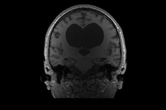
E.S.’s atypical brain may stem from an assortment of issues, including an arachnoid cyst, a fluid-filled sac that forms between the brain and one of its membranes. Hydrocephalus, when excess fluid collects in the middle parts of the brain, may also have compressed E.S.’s brain tissues. On top of that, E.S. is missing most of the tissue that connects the left and right hemispheres, a condition called agenesis of the corpus callosum. In their daily life, E.S. is a professor and researcher in the field of speech-language pathology. COURTESY OF THE FEDORENKO LAB -
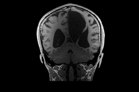
Hydrocephalus has left its mark in K.V.’s brain, pushing neural tissue toward the sides of their skull and pressing the cortex thin. K.V.’s ventricles, cavities filled with cerebrospinal fluid (center), are larger than most people’s and form a fluid-filled void in the center of the brain. K.V. makes their living as a writer. COURTESY OF THE FEDORENKO LAB -
Scientists think that G.C. may have developed a cyst in their left hemisphere early in life. That could have compressed their left frontal, temporal and parietal lobes. Though G.C.’s language areas are squished, the cyst does not seem to have affected their daily life. G.C. is a speech-language pathologist. COURTESY OF THE FEDORENKO LAB
But there’s still a universe of more questions, Fedorenko says. She’s curious if brain functions can overlap, sharing the same cortical machinery in an atypical brain when they might otherwise have set up shop in different locations. And a damaged left hemisphere doesn’t always mean language moves to the right. Sometimes language function stays behind, surviving on the fringes of the damaged region, Fedorenko says. “Nobody knows why that happens.”
Carlson and colleagues reported this kind of adaptation in 2020 in Pediatric Neurology. The team was studying young stroke patients who had perinatal strokes that hit the motor cortex. Carlson says the Interesting Brains Project is valuable because it could tell scientists more about plasticity in the brain at the individual level — how a specific person’s brain has adapted to injury.
Not every person’s brain is able to bounce back. What scientists learn from the project, along with individualized neuroimaging, could help with prognosis, and potentially rehabilitation. “Perhaps if we can tailor intervention options to an individual brain, they might be more effective,” Carlson says.
Getting results will take time. Fedorenko’s team is currently juggling experimental logistics, including scanning a new participant every one to two weeks, performing behavioral tests and analyzing data. Still, they’re seeing some interesting results, Fedorenko says, and hope to submit a paper this summer.
She hopes the project can showcase the range of solutions our brains can, in some cases, employ to deal with a slow or sudden loss of neural real estate. Maybe, she says, the project’s findings will help more people understand “how different you can be and still grow up and do amazing things.”
What is a normal brain anyway?
In an opening note in Fedorenko’s 2022 paper in Neuropsychologia, Elyse wrote about how her brain’s structure doesn’t define her. “Please do not call my brain abnormal, that creeps me out,” she wrote. “My brain is atypical. If not for accidently finding these differences, no one would pick me out of a crowd as likely to have these, or any other differences that make me unique.”
Elyse hopes the message comes through for doctors and research scientists. “I want them to understand that this is a person they’re reading a paper about, not a disembodied brain in a jar,” she says.
One thing Elyse likes about working with Fedorenko’s team is that the research feels collaborative. Scientists rely on close partnerships like this to understand how the brain works under typical situations and how it may recover from injury, says Lesley Fellows, a neurologist at McGill University in Montreal who studies how brain damage affects decision making. People with atypical brains “can give us all kinds of great ideas we might not have thought about,” she says. “They have a unique vantage point.”
Elyse, for example, experiences smell hallucinations. She picks up whiffs of electrical fires whenever she’s under a lot of stress. “When I was in grad school, I would smell electrical fires three times a week,” she says. Elyse hasn’t yet explored this brain quirk with Fedorenko and her colleagues, but she’s open to their ideas for future investigations.
“I want them to understand that this is a person they’re reading a paper about, not a disembodied brain in a jar.”
Elyse G.
For the team’s most recent study, reported in a preprint this year, Elyse, Martha and another sister (one with an “ordinary” brain) participated in hearing tasks inside the MRI tube. Fedorenko’s team wanted to find out how the left or right auditory cortex works when the other side is missing.
You might think that the remaining auditory cortex would have to be enhanced somehow to pull double duty, perhaps taking up extra space, says Tamar Regev, a cognitive neuroscientist in Fedorenko’s lab. But that’s not what the team found.
In both Elyse’s and Martha’s brains, “activity looks completely neurotypical,” Regev says. That suggests there’s some redundancy to the brain’s auditory system, and that the development of one auditory cortex does not depend on the existence of the other.
Elyse is curious what other insights Fedorenko’s team will glean from her brain, and the brains of fellow Interesting Brains Project participants. “My brain is special, unique and interesting,” she wrote in the 2022 paper, “and I am excited that it can help neuroscientists understand the plasticity of the human brain.”