Cells cram DNA into the nucleus in two distinct ways
Some chromosomes look like crumpled balls while others resemble flat sheets of paper, heat maps show
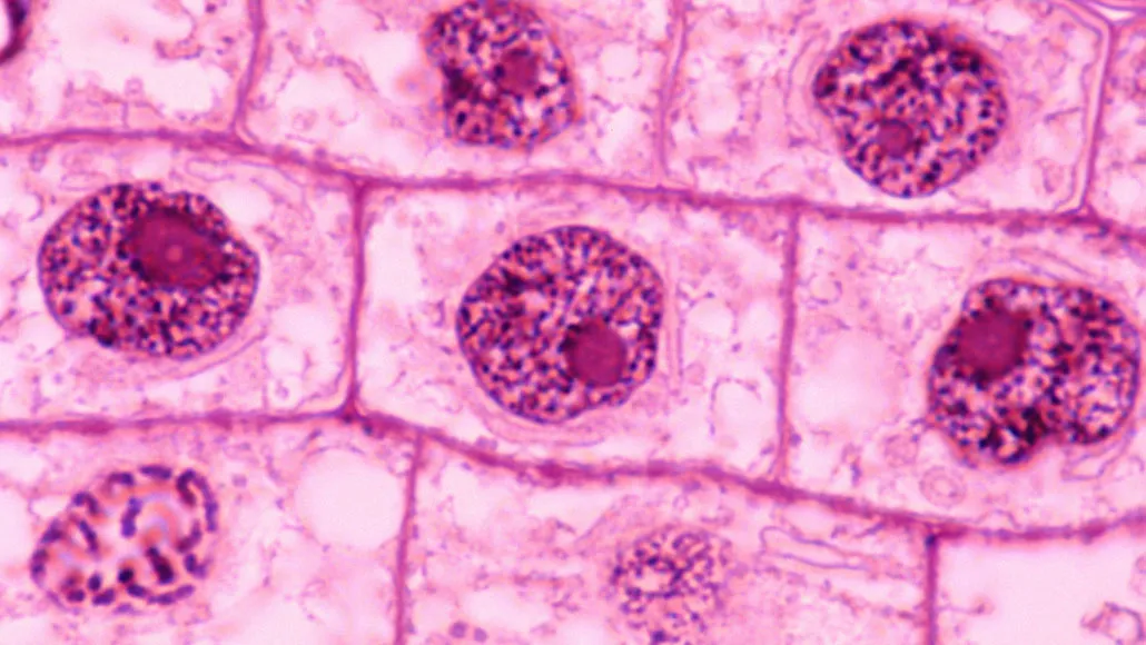
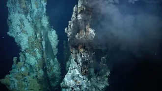
Cramming meters-long DNA strands into a cell’s tiny nucleus is an astonishing feat. A new study suggests that cells from plants (shown), animals and fungi accomplish it one of two ways.
Ed Reschke/Stone/Getty Images Plus
There are only so many ways to cram DNA into a cell’s nucleus, a study suggests.
A cell’s complete genetic blueprint, or genome, is densely packed into chromosomes, condensing meters of DNA into a minuscule cellular vessel only micrometers wide (SN: 8/24/15). But how chromosomes fold to fit inside the nuclei of diverse species is unclear.
There appear to be two methods to stuff all of that DNA in, researchers report in the May 28 Science. Cells can even flip-flop which arrangement they have by inactivating a molecule called condensin II, the team found.
If chromosomes were pieces of paper, some, like those of humans, would look like a crumpled ball inside the nucleus, says Claire Hoencamp, a molecular biologist at the Netherlands Cancer Institute in Amsterdam (SN: 10/8/09). Others, like those of fruit flies (Drosophila melanogaster), resemble flat sheets of stacked paper.
In the new study, Hoencamp and colleagues created heat maps that analyzed how chromosomes in the nuclei of 24 animal, plant and fungal species interacted inside their respective cells. The maps show the average number of connections among chromosomes in a cell’s nucleus — revealing how the genetic molecules fold — “on the scale from white to red,” says Olga Dudchenko, a genomicist at Baylor College of Medicine in Houston. “The more red, the more interactions. The less red, the less interactions.”
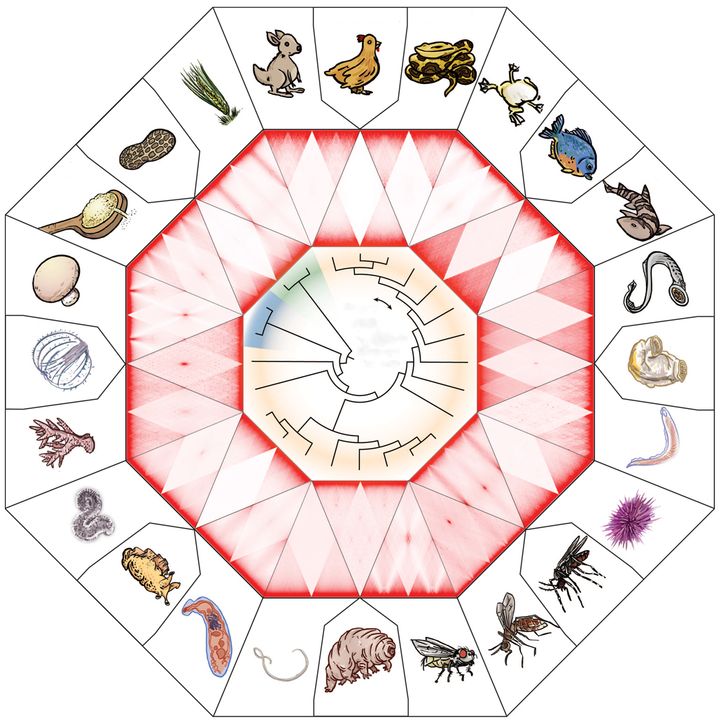
Chromosome contacts
A wide range of animal (colored yellow on the evolutionary tree at center), plant (green) and fungal (blue) species package chromosomes inside the cell nucleus one of two ways, based on how the chromosomes interact inside a nucleus. Heat maps here show the average number of interactions among chromosomes (darker shades of red indicate more interactions). Maps that have large, dark red triangles (such as for the red piranha, top right) indicate chromosomes that crumple up like a wad of paper. Other more checkerboard patterns (such as for the ground peanut, top left) give rise to chromosomes that fold more neatly, like stacked sheets of paper.
Patterns of chromosome interactions for 24 plants and animals
Throughout evolutionary history, organisms across the tree of life have switched among different packing methods, the researchers found. “We worked with a zoo of species, and [at first] it looked like a zoo of patterns of genome folding,” Dudchenko says. “Some maps would look like a checkerboard pattern. Other ones would look like a mattress with weird x’s.” Over time, it became clear that many of the same chromosome folding features were popping up again and again in different species.
Three types of interactions result in stacked sheets of chromosomes, giving the heat maps that checkerboard or mattress look. In one interaction, seen in the ground peanut (Arachis hypogaea) for example, the ends of different chromosomes tend to touch. In another, chromosomes from organisms like fruit flies touch in the middle. And in an interaction seen in bread wheat (Triticum aestivum), the arms of different chromosomes fold on top of one another.
Crumpled ball-like chromosomes, like those of the red piranha (Pygocentrus nattereri), sport a fourth type of interaction. In those structures, a chromosome folds in on itself in a tangle rather than touching other chromosomes, resulting in large red squares on the heat maps.
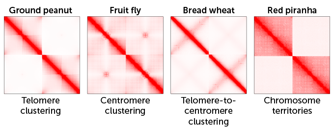
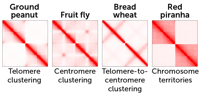
DNA dealings
Chromosomes interact in four different ways, resulting in two types of folding patterns. These selected heat maps for four species show the average number of interactions between two chromosomes; darker shades of red indicate more interactions. Three types of interactions — telomere clustering (ends of the two chromosomes touch), centromere clustering (middles touch) and telomere-to-centromere clustering (arms fold over each other) — make chromosomes fold like flat sheets of stacked paper. Interactions dubbed chromosome territories make chromosomes appear as crumpled balls: In this case, each chromosome is more likely to fold in on itself in a tangle, giving the appearance in the heat map of large, red squares.
Breaking parts of condensin II — a complex of proteins that helps assemble chromosomes as cells divide — can switch which organization a nucleus has. Tweaks to condensin II can make a crumpled human nucleus look like a folded fly’s nucleus, the team found. But some organisms have stacked sheets despite having intact condensin II. That means there may be other factors researchers haven’t yet found that push cells to cram chromosomes into the nucleus in a specific way, Hoencamp says.
We summarize the week's scientific breakthroughs every Thursday.