Coloring the body
Researchers have designed tiny, tunable magnets that may lead to color-coded MRI scans
Micromagnets that finely control radio signals during MRI may one day be used to create “color” scans more sensitive than existing MRI technology. The tiny magnets may also eventually help detect cancer by tagging malignant cells with different colors.
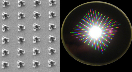
Researchers created tunable magnetic particles, between 2 and 10 micrometers in diameter, which consisted of two metal disks separated by non-magnetic spacers. Changing the size of the spacing between the two disks can precisely adjust the magnetic field generated by an MRI machine. The changes in the magnetic field alter the radio frequency wavelengths sent back to the computer to form an image. These radio signals can then be mapped to different colors, scientists at the National Institute of Standards and Technology Laboratories in Boulder, Colo., report in the June 19 Nature.
“It’s a very innovative paper,” says Chien Ho, the director of the PittsburghNMRCenter for Biomedical Research, who was not involved in the study.
In the future, MRI scans could distinguish between cancerous and healthy cells by coating different-sized micromagnets with substances that bind preferentially to each type of tissue, says lead author Gary Zabow, a physicist at NIST. For example, particles tuned to the show up blue on an MRI could bind to healthy tissue, while particles tuned to appear red could stick to cancer cells, he says.
The technique also increases the sensitivity of MRI, and in the future may allow researchers to detect individual cells, Zabow says. In order for that to be possible, the signal from a single micromagnet bound to one cell would need to be detected.
Currently, MRI images are black and white, although functional MRI often color codes regions of different grayscale intensities to enhance contrast. MRI scans are also not very sensitive. But, the scans can view the body in 3-D and can penetrate deeper into tissue than optical techniques, Ho says.
Before color MRIs become the norm, the researchers need to test the particles in animals, Ho says. As a proof-of-concept, the team used nickel, which is toxic if ingested, to create the micromagnets, although Zabow says less dangerous materials could easily be used. Still, Ho says, it’s uncertain how safe they could make the technology.
“The key question is, when they apply it to biological systems and live animals, what will they get?” Ho says.