Newborn brain has to learn how to feed itself

In infants, blood flow scans used for brain imaging may not reveal nerve activity, study suggests

mouse nerve cell stimulation

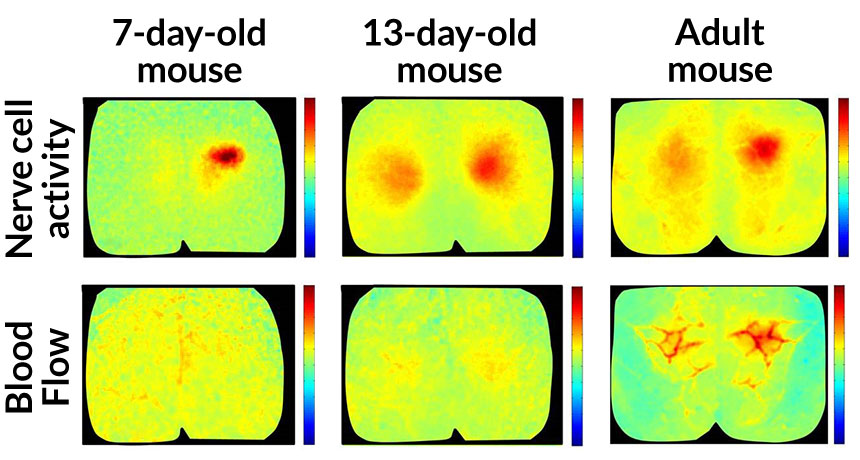
MISS MATCH  After paw stimulation, nerve cells in the brain of a 7-day-old mice become active (top row, left), but blood doesn’t show up (bottom row, left). A 13-day-old mouse has larger nerve cell responses (top row, middle), but still no blood — that doesn’t appear until adulthood (bottom row, right). Red signals higher nerve cell activity and blood flow. 

Hillman Lab/Columbia University’s Mortimer B. Zuckerman Mind Brain Behavior Institute

Busy nerve cells in the brain are hungry and beckon oxygen-rich blood to replenish themselves. But active nerve cells in newborn mouse brains can’t yet make this request, and their silence leaves them hungry, scientists report June 22 in the Journal of Neuroscience.

Instead of being a dismal starvation diet, this lean time may actually spur the brain to develop properly. The new results, though, muddy the interpretation of the brain imaging technique called functional MRI when it is used on infants.

Most people assume that all busy nerve cells, or neurons, signal nearby blood vessels to replenish themselves. But there were hints from fMRI studies of young children that their brains don’t always follow this rule. “The newborn brain is doing something weird,” says study coauthor Elizabeth Hillman of Columbia University.

That weirdness, she suspected, might be explained by an immature communication system in young brains. To find out, she and her colleagues looked for neuron-blood connections in mice as they grew. “What we’re trying to do is create a road map for what we think you actually should see,” Hillman says.

When 7-day-old mice were touched on their hind paws, a small group of neurons in the brain responded instantly, firing off messages in a flurry of activity. Despite this action, no fresh blood arrived, the team found. By 13 days, the nerve cell reaction got bigger, spreading across a wider stretch of the brain. Still the blood didn’t come. But by the time the mice reached adulthood, neural activity prompted an influx of blood. The results show that young mouse brains lack the ability to send blood to busy neurons, a skill that influences how the brain operates (SN: 11/14/15, p. 22).

That finding was enabled by technology that allowed the researchers to see neural activity and blood flow at the exact same time. It’s “a powerful application of cutting-edge imaging techniques,” says neuroscientist Alan Jasanoff of MIT.

Showing that oxygen demands are unheeded during early development is interesting, says neuroscientist Matthew Colonnese of George Washington University School of Medicine and Health Sciences in Washington, D.C. More studies are needed to say whether human infant brains behave similarly and, if so, how this process might sculpt the brain. 

The results don’t mean that fMRI data from young children aren’t valuable, Hillman says. “What we are begging people to do is to make room for this hypothesis, and actually treat it as an opportunity.” Blood flow data might not be a good proxy for neural activity in newborns, but “it may well be measuring a change that is very important to normal brain development,” she says. 

Laura Sanders is the neuroscience writer. She holds a Ph.D. in molecular biology from the University of Southern California.

More Stories from Science News on Neuroscience

From the Nature Index

Paid Content