CT scans show first X-rayed mummy in new light
Images reveal how far radiography has come in 120 years
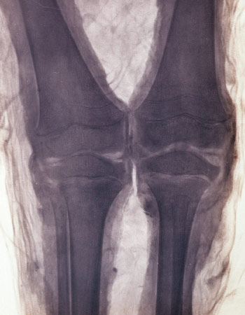
X-rays were the iPhone 7 of the 1890s. Months after X-rays were discovered in late 1895, German physicist Walter Koenig put the latest in tech gadgetry to the test by scanning 14 objects, including the mummified remains of an ancient Egyptian child. Koenig’s image of the child’s knees represented the first radiographic investigation of a mummy.
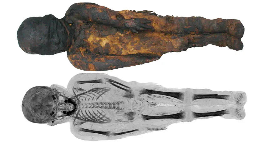
At the time, details on the mummy itself were scant. Originally collected by explorer-naturalist Eduard Rueppell in 1817, the specimen lacked any sort of decoration that might link it to a particular dynasty or time period. Koenig’s X-ray image of the mummy served less to fill in any of those blanks and more to demonstrate the technology’s potential. Since then, radiographic images have revealed hidden artifacts, elucidated embalming techniques and even pinpointed health issues and diseases in mummies.
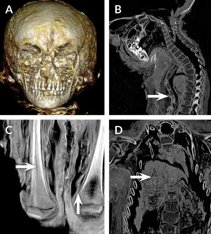
Now, biological anthropologist and Egyptologist Stephanie Zesch of the Reiss Engelhorn Museum in Mannheim, Germany, and colleagues have examined the mummy with modern imaging techniques. CT scans show that the child was a boy. His teeth suggest that he was 4 to 5 years old when he died. Radiocarbon dating places him in the Ptolemaic period, between 378 and 235 B.C., the researchers report online July 22 in the European Journal of Radiology Open.