Famous brain surgery patient H.M. retained a chunk of hippocampus
Amnesia probably due to the loss of other regions, connections
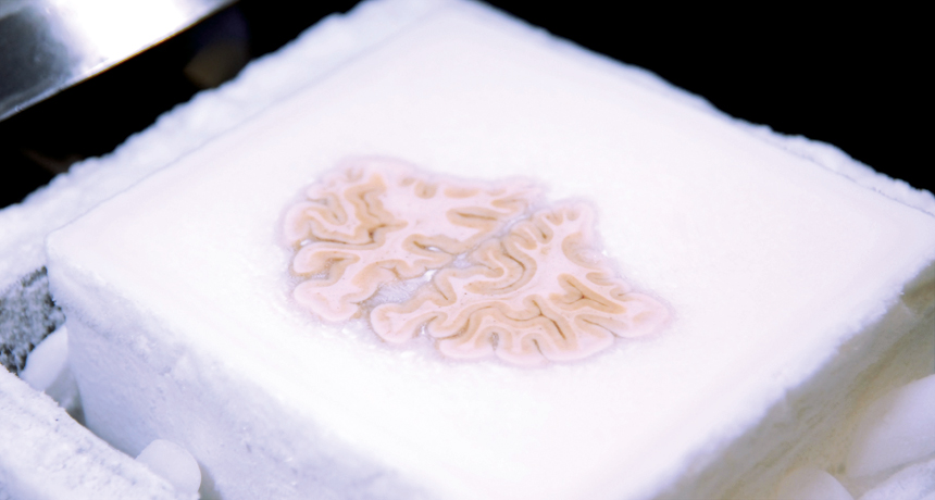
MEMORIES ON ICE Embedded in a frozen gelatin block, Henry Molaison’s brain is about to be sliced and imaged.
J. Annese
After a brain surgeon accidentally took away his memory in 1953, 27-year-old Henry Molaison became one of the most informative cases in psychology. For the rest of his life, the patient known as H.M. donated time to scientists who used the tragedy to study his amnesia. Now, five years after his death, Molaison is still teaching scientists about how the brain makes and stores memories.
A detailed analysis of Molaison’s postmortem brain, generously donated to scientists, reveals a surprising amount of brain tissue in the memory-forming structure known as the hippocampus, scientists report January 28 in Nature Communications. This result shows that Molaison’s severe deficits can’t be pinned exclusively on the loss of the hippocampus, as some scientists previously thought, says neuroscientist Howard Eichenbaum of Boston University, who wasn’t involved in the study.
To ease Molaison’s frequent and severe seizures, surgeon William Beecher Scoville attempted to remove parts of the medial temporal lobe, which includes the hippocampus and other nearby regions. The procedure calmed the seizures but left Molaison largely unable to form new memories. As scientists realized they could link Molaison’s impairments to his drastically altered brain, patient H.M. began to show up in a growing number of research papers.
“The studies on H.M. began the present era of research on how the brain supports memory,” Eichenbaum says.
Because of Molaison’s importance to memory research, Jacopo Annese of the Brain Observatory at the University of California, San Diego and colleagues decided to study Molaison’s brain after his death using a technique that provides a level of detail surpassing methods used on living brains.
About a year after Molaison died, his brain, fixed in formalin, was flown to the Brain Observatory. There, scientists froze it, sliced it into 2,401 paper-thin wisps and photographed them. With these images, Annese and colleagues created a 3-D model of Molaison’s brain.
Scoville’s postsurgical drawings indicated that the entire hippocampus and nearby structures on both sides of the brain had been removed. The new 3-D model showed otherwise.
Molaison retained a surprisingly large amount of what appeared to be healthy hippocampus, the team found. The lingering tissue was the back end of the hippocampus, where the structure curved up toward the top of the head. Scoville may have missed this bit because the suction tube he used to remove brain tissue didn’t follow the bend, Annese proposes. MRI scans from when Molaison was alive revealed some hippocampal tissue, but not as much as the current study finds. Those scans lacked the anatomical precision of the newer method, Annese says.
Because Molaison retained some hippocampus and yet suffered from severe amnesia, other brain structures were probably needed for his memory, Eichenbaum says. Molaison lacked a brain region called the entorhinal cortex, which serves as a signal relay station between the hippocampus and the rest of the brain. The missing entorhinal cortex probably had a big role in Molaison’s impairment, Eichenbaum says.
Comparing Molaison’s memory deficits with those of people known to lack just the hippocampus might help scientists tease apart the jobs of the various brain structures involved in memory, Eichenbaum says.