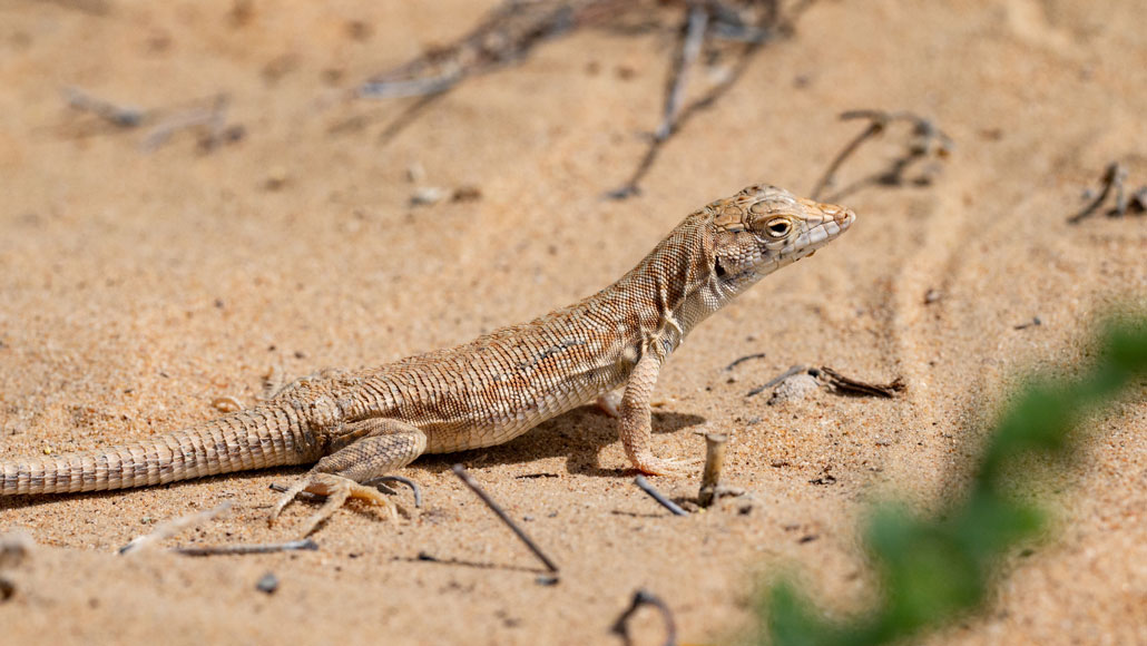
Losing a tail is not ideal, but getting eaten by a predator is even worse. Lizards, like the Schmidt's fringe-fingered lizard shown here, depend on a complex internal structure to help them keep their tails until it’s time to lose them.
John Pereira/inaturalist.org (CC-BY-NC)
Lizards are famous for losing their tails, but perhaps the bigger question should be: How do their tails stay on? The answer may lie in the appendage’s internal design. A structure of prongs, micropillars and nanopores holds a lizard’s tail on tight enough to handle most jarring while remaining primed to drop the tail in case of emergency, researchers report in the Feb. 18 Science.
Self-amputation, or autotomy, of a limb is a common defense strategy in the animal kingdom, including for many lizard species (SN: 3/8/21). But it’s a risky plan: A detachable limb brings with it increased risk of accidental loss from small bumps and snags. “It has to find the just right amount of attachment, so it doesn’t come off easily. But it should also come off whenever it’s needed,” says Yong-Ak Song, a bioengineer at New York University Abu Dhabi in the United Arab Emirates. “It’s a fine balance.”
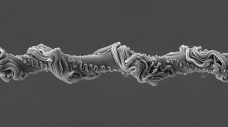
A lizard’s tail consists of a series of segments that connect in a row like plugs into sockets. The tail can break off along any of these points, called fracture planes, depending on how much of the tail the lizard needs to sacrifice. Between each segment, the prongs — eight cone-shaped bundles of muscles arranged in a circle — fit neatly into corresponding sockets, consisting of relatively smooth walls. Each prong is in turn covered in a forest of protrusions, or micropillars, that resemble tiny mushrooms.
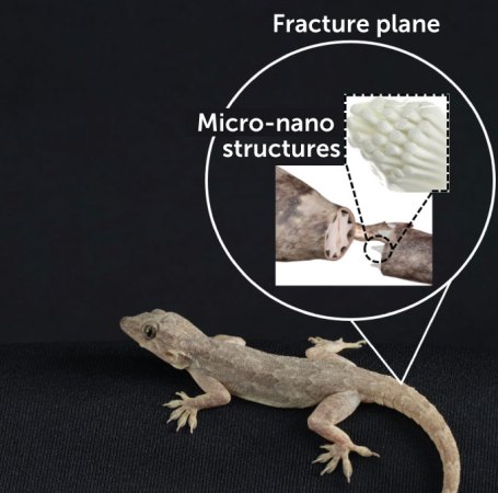
To uncover the function of this structure, Song and colleagues first amputated tails from three species of lizards with a gentle tug and then analyzed the broken appendages under a scanning electron microscope. Zooming in on the mushroom-like protuberances revealed that each one is pockmarked with holes, or nanopores.
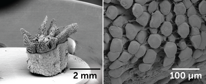
The researchers also noticed slight imprints in the interior walls of the socket left behind by the prong’s micropillars, like fingers pressed lightly into clay. This came as a surprise: They expected that the micropillars would fully interlock within the socket, more like Velcro. Instead, the pockmarked micropillars weren’t providing any extra grip that would secure the tail to its owner.
Suspecting that the nanopore-speckled micropillars must play another role, the team built a replica lizard tail from polydimethylsiloxane, a rubbery, fleshlike material, to mimic the separation of tail from body. This allowed the researchers to examine the forces at work during a tail amputation. They found that the deep crevasses between micropillars, along with the smaller potholes on the micropillars’ surfaces, slow the spread of an initial fracture.
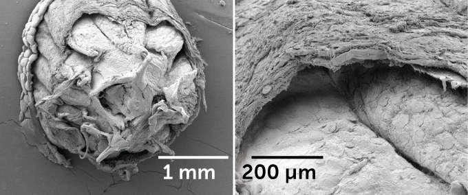
“If there’s a crack coming in and meets a pore, which is a void, then the crack is stopped, and then it loses energy to propagate,” Song says. In other words, the beginning of a fracture can be stopped in its tracks. Every indent and groove helps: The micropillars with nanopores enhanced adhesion 15 times more than smooth prongs without micropillars, and slightly more than micropillars without nanopores. The hierarchical structure of prong, pillar and pore achieves a balance that Song describes as a beautiful example of the Goldilocks principle: not too tight, not too loose.
This adaptation is important for lizards to optimize their survival. While autotomy helps keep a lizard from becoming lunch, it’s a costly defense mechanism that affects a lizard’s ability to run, leap, mate and escape future predators (SN: 1/5/12). So, it’s important that the lizard abandons its limb only when necessary.
This intricately designed system is a perfect example of how evolution can continually work on something to make it more effective, says Bill Bateman, a behavioral ecologist at Curtin University in Perth, Australia, who was not part of the research. “It just blows me away.”
Sign up for our newsletter
We summarize the week's scientific breakthroughs every Thursday.
