Proteins in the Stretch
Tugging at single molecules reveals their secrets
Imagine that one day, instead of folding your clean laundry, you just dump it on your bed and, to your amazement, your shirts, sheets, and towels start folding themselves on their own. A minute later, the disorderly mound has turned into a neat pile of perfectly folded items. Don’t expect to see this outside of a Harry Potter movie, but something just as impressive happens all the time inside your cells. Long, chainlike protein molecules fold up spontaneously and flawlessly into predetermined shapes. There are thousands of kinds of protein in the human body, each with a unique shape vital to its task. Without the power of protein folding, activity in any cell would quickly come to a halt.
The ability to fold into the right shapes is one of many protein properties that remain mysterious. Because proteins are far smaller than the most diminutive bacterium, figuring out what each one looks like in its final, folded form is far from easy. Scientists make a crystal out of many copies of a protein, then shine X rays on it to pick up subtle clues to the protein’s structure.
Scientists have managed to learn the structure of many proteins using such X-ray crystallography, but shape gives only hints of how a protein behaves. To get a better understanding of proteins in action, researchers have recently developed a technique in which they use an atomic-force microscope (AFM) to measure the reaction of individual molecules to applied forces. They call the new method “force spectroscopy” because it reveals information about a molecule by measuring its response to a spectrum of forces.
The technique opens a new dimension of study for proteins, says biophysicist Matthias Rief of the Technology University of Munich. “Similar to X-ray crystallography, which has given eyes to the scientist to see molecules, force spectroscopy provides us with hands to feel them,” he says.
Using the AFM, biophysicists are finally answering basic and longstanding questions about proteins, from how they fold to how they break. The findings have revealed secrets of muscle strength and may contribute to understanding of diseases such as Alzheimer’s and mad cow disease.
A pinch, a pull
The molecule called titin is a giant among proteins. Titins run along muscle fibers, connecting one segment to the next. The largest protein known, titin weighs in at 3 million atomic mass units, or 3 million times the weight of a hydrogen atom. Titin’s weight is 60 times as great as the average for a protein.
Titin’s size made it an attractive target for experimenters working to tease apart individual protein molecules to understand how they work. In 1997, Rief’s research group used an AFM to grasp onto and forcibly unfold individual molecules of titin. A team led by Miklós S.Z. Kellermayer, now at the Howard Hughes Institute in Chevy Chase, Md., simultaneously published a similar achievement with titin. These were the first examples of such a feat being performed on a protein.
The technique depended on the extraordinary powers of the AFM, which was devised originally to make images of the tiniest things by passing a tip over them and feeling for bumps and depressions. The Munich researchers used the same type of microscope for plucking at individual titin molecules and measuring the strengths of their various parts.
The team began by immersing a gold plate in a solution containing normally folded molecules of the protein, some of which spontaneously formed a chemical bond between one of their ends and the surface of the plate. Then, using the tip of the AFM, the experimenters started fishing for the free ends of the molecules.
The business end of an AFM has an arm that, like a diving board, can flex up or down. It bends in that manner when its tip is repelled or attracted by atomic forces in the sample it’s probing. A laser beam bounced off the arm goes in slightly different directions depending on how much the arm is bent. By measuring the deflection of the laser beam, researchers can precisely determine the forces acting on the microscope tip.
Held in a dipped position near the surface of the gold plate, the tip occasionally attaches to the free end of a titin molecule. As the researchers gently move the tip away with a measured force, the deflection of the laser beam continuously indicates how much resistance the protein is putting up against unraveling its structure.
An undisturbed titin molecule resembles a string with tangles regularly spaced along its length. As the researchers gradually increased the force on the protein, it would at first hardly lengthen at all. Then, the tension would suddenly drop, signaling that one of the tangled parts of the protein had just unraveled. This happened several times until the protein was completely unfolded.
When the scientists let the tip sink back toward the gold surface, the protein would fold back up. When they stretched it out again, the characteristic sawtooth pattern of tension changes repeated. Rief and his colleagues were thrilled.
“It was clear that this was an entrance into a completely new world,” says Hermann E. Gaub of Ludwig Maximilians University in Munich, who was part of Rief’s 1997 team.
This first successful measuring of an unfolding protein revealed some interesting properties. Titin can stretch to become 1 micron longer—an enormous distance for a single protein—and not only avoid breaking but also continue to pull back with a significant counterforce. These properties probably endow muscles with their ability to stretch without snapping, says Gaub.
Achilles’ heel
Julio M. Fernandez, now at Columbia University, was also a member of the titin-probing team in Germany. “I came back to the United States, and I totally shifted my research into this,” he says.
At Columbia, Fernandez continued experimenting with titin for a while and then moved on to other proteins. One of those is a small protein called ubiquitin, which latches on to other proteins to signal to a cell that the marked protein should be destroyed. With Mariano Carrion-Vazquez, also of Columbia, and others, Fernandez discovered a hidden weakness by which ubiquitin can be unfolded with much less force than by simply pulling on one of the molecule’s two ends.
Using the technique previously applied in the titin experiments, Fernandez pulled on one end of a gold-anchored ubiquitin molecule. But this time, he also tried pulling on a loop near a molecule’s middle and was surprised to find that the molecule unraveled with less than half the force needed in an end-to-end pull. The loop is “like an Achilles’ heel,” says Fernandez. The team published the results in 2003.
Another group, led by David J. Brockwell of the University of Leeds in England, simultaneously published a similar trick for unraveling part of a different protein. Called E2p, it’s critical to energy processing by the bacterium Escherichia coli.
Fernandez says the two teams’ discoveries may have opened a window onto one way that cells save energy. When no longer needed, proteins are unraveled and dismantled for spare parts at recycling centers inside cells. The recycling machinery may break down proteins at the weak spots that scientists have now identified.
No two alike
Unraveling a protein using an AFM produces sudden, dramatic changes in the microscope’s signal as folds give way. Folding, on the other hand, is a subtler process that’s harder to track. But it’s the folding process—and the mistakes that can happen along the way—that is of most interest to biologists. Again using ubiquitin, Fernandez recently obtained the first view of a single protein as it underwent the folding process from start to finish.
Fernandez stretched out a ubiquitin molecule with the AFM tip, but this time, he took readings as the tip moved back toward the gold surface. An apparatus more sensitive than that in the protein-unfolding experiment tracked the decreasing distance between the two ends as the ubiquitin protein cinched back up into its natural form. The readings showed the protein’s length fluctuated as it rearranged itself.
Despite decades of folding research, “no one had ever seen the folding trajectory of a single protein before,” Fernandez says.
A common view among protein researchers had been that proteins always follow the same folding path. Fernandez, however, observed that the trace of how hard the folding ubiquitin molecule pulled on the microscope tip was different each time that he ran the experiment. He concludes that there are many ways in which ubiquitin can fold into its natural form.
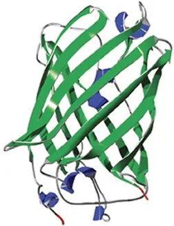
Once a protein has folded properly, what makes it stay compact? In other words, what makes a protein stable? Rief and Hendrik Dietz, also of the University of Munich, have been investigating this question by studying a molecule called green fluorescent protein, which was originally isolated from glowing jellyfish. Genetic engineers have prompted many other organisms to produce the protein. The resulting glow in all or part of a plant or an animal is a common marker in genetics studies.
Scientists still don’t understand how the structure of the protein enables it to glow. Although Dietz and Rief haven’t yet answered that question, they have discovered a critical element of the protein’s stability.
The main structure of green fluorescent protein, when properly folded, looks like a barrel. Dietz and Rief found that by using an AFM to pull on just one small coil, called an alpha helix, at the edge of this barrel, they can make the protein highly unstable. A tug on the protein made it lengthen by 3 nanometers, just the amount one would expect when something the size of an alpha helix unfolds from the barrel. However, with the alpha helix unfolded, the rest of the molecule spontaneously unfolds.
Says Rief, “The little alpha helix glues [green fluorescent protein] together.”
Dietz and Rief suggest that this result, published in the Nov. 16, 2004 Proceedings of the National Academy of Sciences, may eventually lead to the protein’s use as a force sensor inside cells. Green fluorescent protein could be spliced into structures in a cell that are subject to tension forces. Whenever the tension reached a certain level, the fluorescent protein would unravel, and the green glow would disappear.
Tiny hands
The future looks bright for such single-molecule investigations of proteins. Several teams are looking into the misfolding of proteins, which scientists theorize to be the problem underlying brain illnesses, including Alzheimer’s disease and Huntington’s chorea. In particular, the researchers hope to gain insights about the infectious proteins presumed to cause mad cow disease and the related Creutzfeldt-Jakob disease in people.
Called prions, these molecules are notoriously difficult to study. They all come in two varieties: the normal form, which is harmless, and the infectious form, which causes disease. What’s frustrated researchers so far is that the normal and disease forms of these proteins seem to differ only in the way they’re folded.
The AFM protein-stretching technique might end that frustration. “We think that using this method we could distinguish and better understand the two types of folded shapes [that] prion proteins can assume,” says Harald Janovjak of the University of Technology in Dresden, Germany, whose group is pursuing this type of experiment.
Gaub is optimistic about the potential of the technique for studying proteins in general. “If one is capable of handling a single molecule, a single protein,” he comments, “then all the questions that have to do with the mechanics of molecules, with the folding of molecules, and with their biological function are in your hands.”