An X-ray laser so bright and fast that it puts a paparazzi zoom lens to shame has allowed researchers to snap pictures of celebrity molecules that typically avoid scientists’ prying eyes. The method should prove widely useful for investigating the structure and activity of drugs, molecules for fuels and other materials.
Scientists have now used the new technique, 10 years in the making, to image an important photosynthesis protein and a virus. And the method should eventually allow researchers to make movies of molecules interacting with each other. As the infrastructure of the cellular world, proteins are of particular interest; witnessing them in action could shed light on processes from brain-cell activity to photosynthesis.
“This will be extremely interesting in just about all biological systems,” says physicist Henry Chapman of the Center for Free-Electron Laser Science in Hamburg. Chapman is a member of the two international teams who report the technique’s success February 2 in separate Nature papers. “After all, the reason we want to obtain high-resolution 3-D images of proteins is to work out how they work and what they do.”
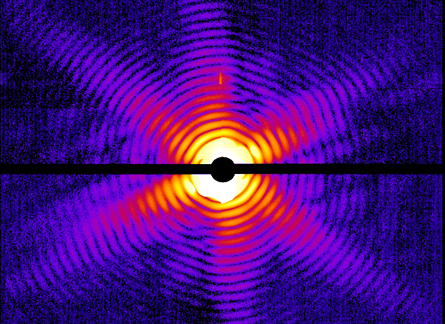
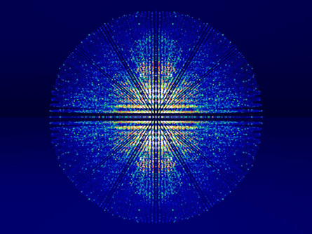
Scientists already use X-rays to image proteins; by collecting the diffraction patterns made when an X-ray strikes a molecule, researchers can piece together its three-dimensional structure. But current techniques require hefty, pure molecules that must be laboriously isolated and crystallized before they can be looked at. Other methods, such as electron microscopy, require extensive prep work such as freezing the sample, and then researchers can look only at slices.
But the X-ray laser used in the new work is so much brighter and faster than its predecessors that researchers don’t need to grow their molecule of interest into a big, sturdy crystal. Eventually, researchers hope, the technique may reveal molecules interacting in their native habitat.
“The biggest problem has been membrane-bound proteins — they are very hard to get a detailed view of,” says biophysicist Sebastian Doniach of Stanford University, who was not involved in the research. “But these are the proteins that are really important for understanding how things enter the cell, how cells such as nerves signal, how drugs interact with a target cell.”
The new method uses the Linac Coherent Light Source, which came online in 2009 at the SLAC National Accelerator Laboratory in Menlo Park, Calif. This free-electron laser produces pulses of what are called hard X-rays that are a billion times brighter than the synchrotron X-rays used in traditional protein crystallography. The laser light’s wavelength is close to the width of an atom, allowing resolution on an atomic scale. And the laser’s pulses are so short that it can capture images with a “shutter speed” on the femtosecond scale, quicker than a trillionth of a second.
By feeding a continuous stream of molecules or another microscopic sample into the X-ray beam, scientists can take snapshot after snapshot, capturing meaningful structural information moments before each particle explodes into oblivion.
“The molecule in the beam doesn’t know what hit it,” says Chapman. “It just disappears in a flash of light.”
Measurements suggest that the samples, be they proteins or virus particles, get hotter than the surface of the sun. The X-rays tear electrons from a sample, which becomes plasma before blowing up in a blaze of glory. But not before a diffraction pattern is captured that reveals how the X-rays scattered off the sample. Combining millions of these snapshots allows reconstruction of the sample’s structure.
In one round of experiments, researchers used a specialized water jet to shoot a stream of tiny crystals of photosystem I, a protein important in photosynthesis, into the X-ray beam. Researchers also shot a stream of aerosolized virus particles to the X-ray beam and successfully created an image of the beastie. Both the protein and the virus had been imaged previously by older techniques, allowing researchers to confirm the resulting structures.
The possibility of “diffraction before destruction” was presented 10 years ago in a Nature paper coauthored by Janos Hajdu of Uppsala University in Sweden. The demonstration of what was once just calculations on a page is thrilling, says Hajdu, a coauthor on both new papers.
“My dream is to see a cell at near-atomic resolution before I die,” Hadju says. “How is this protein folding? What is this repressor doing on this gene? Why is this channel open? Those are things I want to know.”
