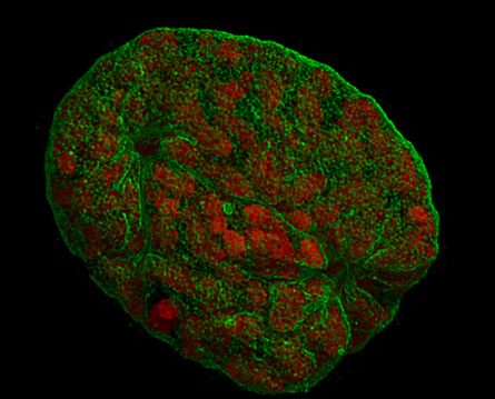
This 3-D view of a mouse cell’s DNA-containing nucleus was taken using a modified light microscope. Researchers stained the cell with normal fluorescent dyes and mounted it on a standard microscope slide. To capture this unprecedented image, the scientists illuminated the cell with three intersecting beams of light and processed the resulting image with a computer. While other 3-D microscopy techniques exist, the new method, called 3-dimensional structured illumination microscopy (3D-SIM), is the first to combine a 3-D view, multiple colors and a resolution of 100 nanometers — twice that of the best conventional light microscopes, the team reports in the June 6 Science. “You can do this 3-D reconstruction with any confocal [light] microscope,” notes research team member Heinrich Leonhardt, a geneticist at LudwigMaximiliansUniversity in Munich, Germany. In the image, red indicates the bundled up DNA inside the nucleus and green shows the nuclear membrane.