Beyond Blood
Next-generation MRI scans offer a sharper picture of the brain's inner workings
The test subject lies on his back, legs stretching from the tunnel-shaped brain scanner. Flat-screen computer monitors fill the cramped control room for the MRI machine. The subject watches one screen, where phrases flash, such as “Jesus is the son of God,” “God puts good and evil everywhere,” and so on.
“We’re doing a study on religion in the brain,” says the technician at the National Institutes of Health (NIH) in Bethesda, Md.
Another monitor reveals a black-and-white cross section of the subject’s brain. The picture refreshes every few seconds. The image is mostly a blur, but some smears appear brighter, others darker. It’s the brain on religion.
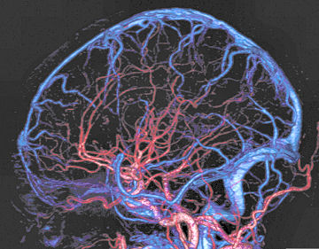
The scanner isn’t taking photos of brain cells contemplating the afterlife. Instead, the snapshots capture blood flowing to the cells. Scientists call this measurement BOLD—short for blood-oxygen level-dependent. More blood equals more thought, the theory goes. Combined with traditional MRI (magnetic resonance imaging), the technique has revolutionized neuroscience, providing tantalizing glimpses into the biology of cognition.
Scientists call this method of scanning a brain at work functional MRI, or fMRI. Today, nearly every fMRI study relies on blood flow.
However, many scientists say that, while useful, blood flow is an indirect gauge of brain activity at best, and misleading at worst. More precise measures of the mind will offer clearer and more complete pictures of human and animal brains at work.
“Using BOLD to study how the brain works is a little bit like feeling different parts of a computer—feeling how hot they are—and trying to understand how it works based on that,” says Alan Jasanoff, a neuroscientist at the Massachusetts Institute of Technology and a leading proponent of the budding field of bloodless MRI. He studies chemicals that brighten the magnetic signal revealed by an MRI scanner. Other researchers hope to tap the minuscule electric currents that flow through neurons, while still others hunt for subtler physiologic changes.
Although still in its infancy—and flush with ideas but short on results—bloodless MRI will someday usher in a sea change in our understanding of the brain, its proponents say. The new techniques could provide more detailed maps of brains, illuminate the connections between distant regions of the brain, and diagnose diseases like Alzheimer’s.
Leaky pipes
Early efforts to map the human brain were as invasive as they were primitive. In the 19th century, scientists linked form to function by dissecting the brains of deceased patients. Surgery offered another window into the mind, as physicians prodded brains. Gentler techniques, such as electroencephalography (EEG) and positron emission tomography (PET) emerged, but suffer other shortcomings. PET releases radiation that is potentially dangerous, and EEG only picks up superficial waves.
Functional MRI seemed like the perfect solution. It is noninvasive and measures brain activity via blood flow with the same machine that diagnoses a badly sprained ankle. The scanner generates an enormous magnetic field tuned to physiologic signals—the hemoglobin in blood, for instance. Then, while a test subject performs a task, such as viewing pictures, the MRI scanner records brain activity. Computers translate the whole affair into a 3-D map, indicating which areas lit up during the task and which dimmed.
Invented in the early 1990s, fMRI was slow to catch on. But the technique eventually became a blockbuster among neuroscientists, says the NIH’s Peter Bandettini, an early pioneer who has undergone thousands of scans himself.
In an early experiment, Bandettini alternately tapped his left and right fingers while lying in the scanner. A movie captured the effect: A white smudge, where blood flow increases, jumps between the right and left sides of his motor cortex, a brain region responsible for movement.
For all its usefulness, fMRI measures blood flow, not electricity, the lingua franca of neurons. “The first time I went to an MRI meeting I felt like an electrician in a plumbers’ convention,” says Nikos Logothetis of the Max Planck Institute for Biological Cybernetics in Tübingen, Germany.
Another common gripe against BOLD is the lag between electrical brain activity and blood flow, Jasanoff says. Tap a finger, and neurons in the motor cortex pulse. Blood soon flows through capillaries to the cells in the motor cortex, but the BOLD signal peaks when blood courses through bigger vessels and subsides several seconds after the initial finger tap. This delay means that when several regions of the brain light up in succession, the BOLD signal might mask the order of activation.
“If you think about what you do with your brain, you would agree that 5 seconds is actually an infinity,” Jasanoff says. Imagine driving a car with a 5-second lag between seeing a curve and turning the steering wheel.
The spacing of blood vessels creates another problem with linking blood flow to brain activity. Because vessels form a loose net around tightly packed neurons, researchers have trouble distinguishing signals from brain cells that are less than a millimeter apart.
Perhaps an even bigger drawback of BOLD may be when it lies. Some brain cells, called inhibitory neurons, actually dampen brain activity by squelching other neurons. Yet they generate the same BOLD signal as other neurons.
Search for a signal
In 2006, French neuroscientist Denis Le Bihan and colleagues in Japan published results that continue to reverberate through the small world of brain imaging. Le Bihan’s MRI scanners were tuned to detect water molecules as they moved across the membrane that surrounds brain cells, a technique called diffusion MRI. By scanning the brains of six subjects gazing at a flickering dartboard, his team found a brain signal that peaked a few seconds before blood flow could be detected.
Previous research had suggested that neurons swell ever so slightly when they become active. Le Bihan, director of the Federative Institute of Research on Functional Neuroimaging in Saclay, France, thinks his diffusion MRI experiments captured this swelling.
Yet no one has repeated his results. Some claim that the signal may have been caused by changes in blood flow, not water diffusion. “The result may have been an artifact or one of billions of scientific results that seems to be here one day and gone the next,” Jasanoff says.
Despite skepticism, Le Bihan is determined to prove his results weren’t a fluke. He recently compared diffusion MRI with a technique that gauges brain activity with infrared lasers. The diffusion signal beat out the laser’s measurements, he says.
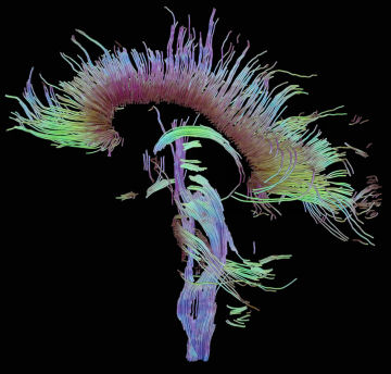
Less controversially, neuroscientists use diffusion MRI not to track activity but to map physical connections between distant regions of the brain. In 2007, a Dutch team studied people who perceive words as colors, one form of a condition called synesthesia. Scans with diffusion MRI suggested that the synesthete brains boast extra connections between a region involved in word and color processing and one linked to consciousness.
Though water diffusion may prove to be a better proxy for brain activity than is blood flow, diffusion MRI remains a circuitous measure. For an even closer pulse on the brain, some scientists are trying to tap into the native tongue of neurons-electricity.
Every time a neuron fires, electrically charged molecules flood in and out of the cell, producing a tiny electrical current. An MRI scanner could likely detect the magnetic field created by the current, says Bandettini.
To test the idea, his team put a petri dish of neurons under a powerful MRI scanner. The cell culture produced a faint signal that disappeared when the researchers added a chemical that blocks the cells from firing. Though the results showed it was feasible to measure the electric fields of neurons, the technological leap from a soup of cells to a human brain isn’t trivial. “It will take some sort of breakthrough for it to work,” Bandettini says.
Another way of doing bloodless MRI is to tweak the magnetic properties of brain cells with chemicals called contrast agents, says Jasanoff. Most of the chemicals have to be injected directly into the brain, and some are toxic, undercutting their usefulness. However, they reveal measures of brain activity—such as changes in pH or calcium levels—that are more substantive than blood flow.
A better contrast agent might be ferritin, a protein complex that sops up free iron, toxic to cells. But ferritin’s natural levels are too low for MRI to pick up. To solve that problem, researchers at Carnegie Mellon University in Pittsburgh infected mouse brain cells with a virus that inserts extra copies of the ferritin genes into the cells. In the future, scientists may sew other genes to the ones for ferritin, creating an MRI signal to match most any cellular chore.
Such tools, though unavailable for human research now, are already making a difference in animal research, says Daniel Turnbull, a neuroscientist at New York University. Using a manganese contrast agent that’s sensitive to brain activity, Turnbull found that mice rewire their brains after hearing a monotonous tone for a day.
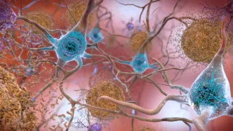
While chemicals like manganese probably won’t fly in humans because of toxicity, researchers haven’t written them off completely. Turnbull’s lab is working on a molecular sensor to detect plaques associated with Alzheimer’s disease. And in 2006, Swedish researchers labeled a nontoxic body breakdown product, pyruvate, with a short-lived magnetic signature. The researchers injected the tagged pyruvate into rats and pigs, and picked up its signal with MRI.
Bold moves
Despite such glimmers of progress, many say measuring blood flow is still the best way to see brains—and will be for some time. By carefully timing fMRI tasks, researchers can detect responses within milliseconds, Bandettini says.
Neuroscientists have also devised new ways to apply conventional brain imaging. Real-time fMRI, where scientists get a live read-out of a subject’s brain, may help treat chronic pain. In one experiment, researchers scanned the brains of pain sufferers and told them what their brains were doing at any instant. Eventually, the patients learned to command a region of the brain linked to pain and even get some relief. Although they sound far-out, mind reading and lie detection may soon become possible with fMRI. On March 5, scientists reported that they deduced the pictures subjects viewed by analyzing the BOLD signal from a region of their brain called the visual cortex (see “Pick a photo, any photo,” in this week’s issue).
“I think it’s much more fruitful to work on BOLD than to completely start over,” Bandettini says.
Other researchers are more optimistic about the prospects for bloodless MRI, eyeing a potentially huge payoff. Today, fMRI does a decent job of identifying regions of the brain associated with particular thoughts or activities, but the network between these regions is largely unmapped, says Allen Song, a neuroscientist at Duke University in Durham, N.C.
“If I pinch you, you feel pain. That’s well understood,” he says. “But from that pain you remember the event. We don’t really have a good handle on what causes all that.” Filling in such missing links is reason enough to pursue new ways of seeing the brain, however speculative, Song says. He is working on a way to image the brain by measuring charged molecules flooding out of active nerve cells.
BOLD continues to be the workhorse of brain imaging, including the NIH efforts to find the seat of religious belief. Jordan Grafman, the neuroscientist leading the study, suspects that the religious messages will light up the prefrontal cortex. Neuroscientists think this region is responsible for many of the cognitive differences between humans and their primate cousins. Yet until scientists perfect new, more direct ways of capturing the human brain at work, researchers may be seeing the effect of religion on blood, not on the brain.