Fallout from nuclear bomb testing presaged today’s radioactive tracers
Excerpt from the February 20, 1965 issue of Science News Letter
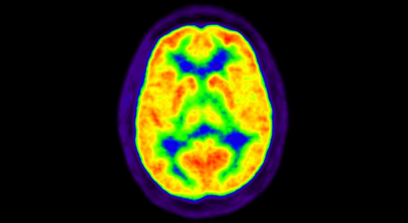
NUCLEAR BRAIN In 1965, researchers developed a way to track radioactive carbon in the body. Today, positron emission tomography (PET) scans work by the same method to diagnose diseases.
TRIUMF Lab/Flickr (CC BY-NC-SA 2.0)
Carbon tra ces changes — Fallout from nuclear bomb tests is allowing University of California at Los Angeles scientists to develop a new method for tracing vital chemical and physical changes in the human body.