Faulty cellular antennae may cause a heart valve disorder
The discovery could help researchers understand how mitral valve prolapse develops

HEART MURMUR Mitral valve prolapse — a leaky valve between two chambers of the heart — can produce a “click” sound that is detectable with a stethoscope.
Rocketclips, Inc/Shutterstock
Cells with faulty antennae that can’t get their signals straight may be behind a common heart valve disorder.
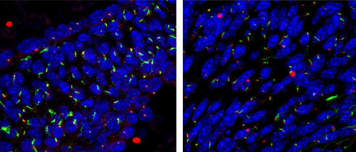
Newborn mice engineered to develop a flawed heart valve had stunted primary cilia in cells that help to form the valve during development, researchers report online May 22 in Science Translational Medicine.
The heart valve disorder, called mitral valve prolapse, “is characterized by significant abnormalities in the composition and organization” of valve tissue, says Joy Lincoln, a heart valve researcher at Nationwide Children’s Hospital in Columbus, Ohio. Those abnormalities compromise the valve’s structure and function. The new work hints that primary cilia play a role in this improper development, she says.
The condition, which affects about 2 percent of the U.S. population, causes the valve separating two heart chambers — the left atrium and the left ventricle — to not seal properly. Normally, when the left ventricle pumps oxygenated blood to the body, the valve closes tightly so blood doesn’t backtrack into the atrium. But with mitral valve prolapse, parts of the valve bulge into the atrium, which can let blood through.
A leaky valve can produce a heart murmur, a “click” sound a doctor can hear with a stethoscope. Some people with the condition don’t experience any symptoms, while others may have rapid heartbeats or discomfort in their chest. If there is a lot of leaking blood, the heart may develop an infection, blood clots may form and raise a person’s risk of stroke or heart attack, or a person’s heart may eventually fail. The only way to fix the valve is with surgery.
Primary cilia, solitary cellular antennae thought to be crucial for signaling between cells (SN: 11/3/12, p. 16), differ from motile cilia, which work as a group to move things along the respiratory or reproductive tracts, for example. Scientists previously thought primary cilia were no longer functional. The idea that primary cilia might be involved in the heart valve’s troubles sprung partly from the recognition that people with polycystic kidney disease develop mitral valve prolapse more often than those in the general population. The kidney disease is one of a number of rare diseases called ciliopathies, which have in common dysfunctional primary cilia.
Russell Norris, a cardiovascular biologist at the Medical University of South Carolina in Charleston, and colleagues identified a multigenerational family with an inherited form of mitral valve prolapse. The affected family members shared a mutated gene linked to primary cilia. This discovery provided the impetus for exploring a possible role for the cilia in the disorder.
Newborn mice engineered to have the mutated gene ended up with undersized primary cilia in cells in their mitral valves during development and showed signs of mitral valve prolapse. Cells with faulty antennae seem to “get confused on where they are supposed to go” and mismanage the building of the valve, Norris says. As a consequence, “the makeup of the valve tissue itself is altered.”
In an analysis of human genetic information, the team also found that small variations in a set of 278 genes tied to primary cilia occurred more frequently in a group of about 1,400 people with mitral valve prolapse, compared with a group of over 2,400 people without the valve disorder.
The team plans to work next on figuring out how these dysfunctional primary cilia, present only during development, actually might cause mitral valve prolapse. Understanding how the disease develops may help doctors figure out the right time to intervene, which isn’t always clear, Norris says.
“The current mode of treatment is to sort of watch and wait,” he says. But repairing a heart valve won’t help a patient if the “heart has already gotten to the point where it’s starting to fail.”