Immune cells’ intense reaction to the coronavirus may lead to pneumonia

The virus appears to target macrophages that reside in the lung tissue

COVID patient lung x-ray checked by doctors

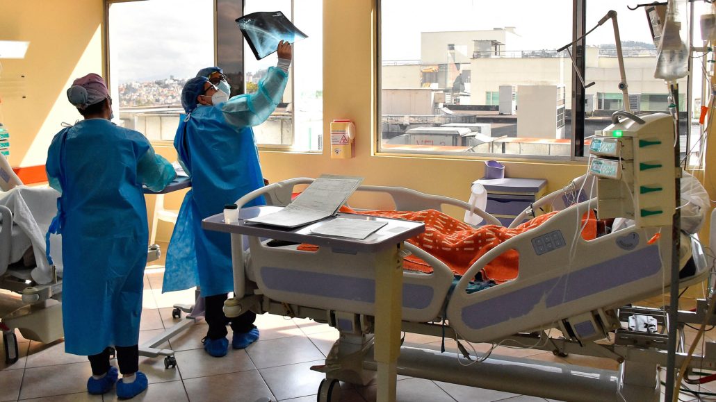
Health workers in Ecuador examine a chest X-ray from a COVID-19 patient in early 2022. A new study suggests that immune cells that reside in the lung tissue may play a role in the progression of a coronavirus infection to pneumonia.

RODRIGO BUENDIA/Getty Images

The intense reaction of one of the lungs’ guardians against infection may help explain why COVID-19 can become severe.

The guardians, immune cells called interstitial macrophages, patrol lung tissue. These cells can be infected in a big way by SARS-CoV-2, researchers report online April 10 in the Journal of Experimental Medicine. Overwhelmed by the virus’ onslaught, the cells’ extreme inflammatory response may contribute to the development of pneumonia, a disease that damages the lungs and makes breathing difficult.

“There is this gap in our knowledge of how human lung tissue really responds in the earliest phases” of a SARS-CoV-2 infection, says José Ordovás–Montañés, an immunologist at Boston Children’s Hospital and Harvard Medical School, who was not involved in the research. The new work has “put a spotlight” on interstitial macrophages’ role, he says. “I think this is an interesting piece of the puzzle.”

A bout of COVID-19 can begin after someone breathes in the coronavirus, which — launched by a sneeze or a cough — spreads through the air (SN: 12/16/21). Evidence suggests the virus first infects cells that line the nasal cavity or the throat, and then the immune system response kicks in. From that point, many people clear the infection, says Catherine Blish, a viral immunologist at Stanford University School of Medicine. But some don’t, and the virus can spread into the airways of the lungs and infect cells lining the air sacs, the structures where oxygen and carbon dioxide are exchanged.

Blish and her colleagues wanted to investigate the next steps of a coronavirus infection in the lungs and what might drive the progression to pneumonia. The team worked with thin slices of human lung tissue, procured from organ donations or surgeries in which some of the tissue needed to be removed. The researchers exposed the tissue to SARS-CoV-2 to see which cells became infected. “It was really no contest,” Blish says. The vast majority were macrophages, immune cells that take up viruses and other pathogens and “eat” them in order to present the parts to other immune cells. This helps activate the body’s immune response.

Specifically, there were two types of macrophages: Those that reside in the lung tissue, called interstitial, and those associated with the air sacs. The researchers took purified macrophages of each type and put them in a dish with SARS-CoV-2 for additional experiments to show that the cells were being infected, not just gobbling up virus. The team also probed the immune response of the two populations of macrophages. Those that reside in the lungs’ air sacs weren’t as dominated by the virus and produced a proportionate inflammatory response. 

But the coronavirus was able to take over the cellular machinery of the interstitial macrophages. The team found that in response, these macrophages ramped up their production of proteins that target viruses and call in other immune cells. The cells are “sending massive alarm signals … ‘There’s an invader, danger, danger’,” Blish says.

In the body, this kind of immune response can lead to a huge influx of cells and inflammatory proteins into the lungs’ air space, Blish says. That can compromise the air sacs’ ability to function and help set the stage for pneumonia.

Blish and colleagues also found that SARS-CoV-2 enters the interstitial macrophages through a different receptor than the very well-known entry point for other cells, ACE2. This may help explain why treatments that target ACE2 haven’t worked well for severe pneumonia, Blish says.

Experiments with lung slices can’t exactly replicate what’s going on in the body. The study doesn’t answer how the virus would get into the lung tissue and gain access to the interstitial macrophages, Blish says.

“It’s a sensible model to put forward,” Ordovás–Montañés says. But with the lung slices, “you give equal chance to any cell that was either on the outside part or on the inside part of the lung” to become infected. It may be helpful to complement this research with studies in animal models in which the lungs’ physiology is intact, he says, although those types of studies also don’t exactly translate to what’s happening in people. “Each model is going to give you a slightly skewed view of reality.”

Aimee Cunningham is the biomedical writer. She has a master’s degree in science journalism from New York University.

More Stories from Science News on Health & Medicine