Laser light made inside cells

Microscopic implants trap, amplify light, letting researchers track cellular activity

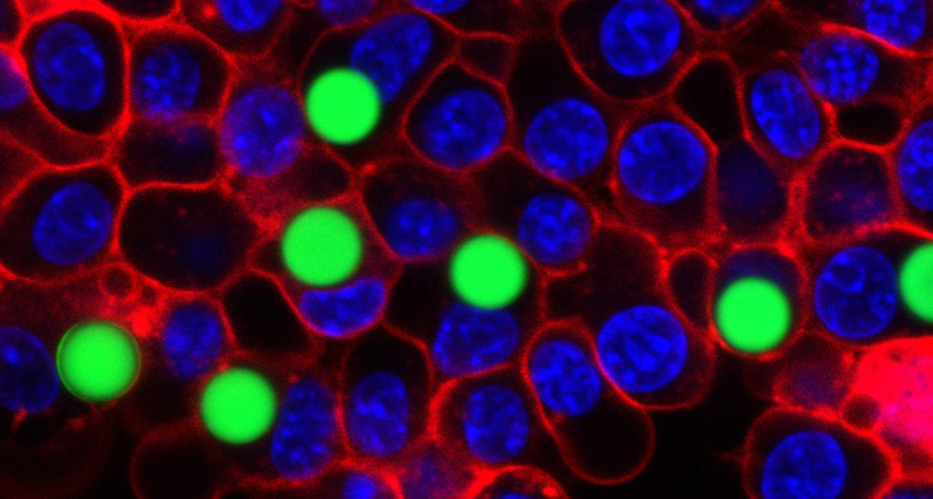
LASER TAGS  Seven of the cells in this micrograph contain lasers in the form of plastic beads (green). The cells are enclosed in red; the nuclei are blue.

Matjaž Humar and Seok-Hyun Yun

Biologists often use lasers to probe cells. Now, for the first time, cells have returned fire.

Harvard University researchers have created intracellular lasers by implanting microscopic beads and oil droplets into animal cells. When energized by an outside laser pulse, an implant traps and amplifies light and then emits a laser pulse of its own. “It’s a wonderful way of coupling optics to cells to learn about biological processes,” says chemist Richard Zare of Stanford University. The microscopic lasers, reported July 27 in Nature Photonics, could allow scientists to track the motion of thousands of individual cells.

The new technique is far from the first to coax cells to generate their own light. Biologists routinely scrutinize cells under the microscope with the help of fluorescent dyes and proteins that glow when energized by an external laser pulse (SN Online: 10/8/14). But those cells emit light with a wide span of wavelengths, says physicist Seok-Hyun Yun. That can make it difficult to separate the cells’ glow from background illumination. Yun and colleague Matjaž Humar set out to implant cells with lasers, which emit light only at specific wavelengths.

RADIATING PIG SKIN Cells located beneath the skin of a pig contain fat globules —similar to the oil droplets that researchers used in experiments — that emit laser light when energized by a pulse from an optical fiber. Matjaž Humar and Seok-Hyun Yun
The lasers had to be small and simple to function unobtrusively inside a cell, so the physicists used either plastic beads or dyed oil droplets. In some ways, these microscopic implants are like fluorescent dyes: Add energy with an external laser pulse and they radiate light. But rather than producing an immediate broad-spectrum glow, the droplets and beads trap light inside. The light circles repeatedly around the orbs’ circumference, each revolution coaxing the production of even more light. After about a nanosecond, millions of photons escape as laser light. “You could see a single cell lasing with the naked eye,” Yun says.

The component wavelengths of the laser light depend on the size of the spheres, Yun says. Humar and Yun showed that they could measure cells’ internal pressure by charting changes in laser light that occur when cellular fluid compresses the oil droplets. The rigid beads, which don’t change in size, could be used to tag individual cells. By using beads that differ in diameter by as little as 2 nanometers, Yun says that scientists can distinguish between thousands of individual cells by their laser signature. 

More Stories from Science News on Life

From the Nature Index

Paid Content