Memory echoes in brain’s sensory terrain
Psychologists have long noted that any of the sights, sounds, and other inputs that make up an experience can, if encountered again, ignite a memory of the event.
In two independent studies, neuroscientists have taken steps to untangle the brain processes that link sensations to memories. When people recall information composed of sights and sounds, neural activity surges in some visual and acoustic areas of their brains just as it does when they first formed the memory, the two teams report in the Sept. 26 Proceedings of the National Academy of Sciences.
These particular brain regions handle sensory information only after it has passed through the separate neural gateways for sight and sound. Parts of the frontal brain, already thought to coordinate memory retrieval, recruit these secondary sensory areas to help assemble memories, both groups theorize.
“These new studies set the stage for more intensive explorations of memory retrieval in the brain,” remarks Anthony D. Wagner of the Massachusetts Institute of Technology in Cambridge.
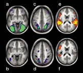
In the first investigation—conducted by Mark E. Wheeler, Steven E. Petersen, and Randy L. Buckner, all at Washington University in St. Louis—18 volunteers intensively studied sets of written labels. Each label was paired with a picture or a sound. For example, the label “dog” was shown with a dog’s picture for nine participants and with the sound of a dog barking for the rest.
The next day, the researchers used a technique known as functional magnetic resonance imaging to measure brain activity under two conditions. In the first, each person reviewed the pairings that they had learned. In the second, they tried to recall the matching pictures or sounds when shown written labels.
Volunteers remembered most of what they had studied. Parts of the visual cortex showed activity boosts when the participants recalled pictures, whereas parts of the acoustic cortex reacted strongly during sound retrieval. Those areas fell within the regions activated during the review.
In the other work, directed by Lars Nyberg of Umeå University in Sweden, previously studied printed words activated parts of a person’s acoustic cortex if the words had been paired with distinct sounds during learning.