One path that fear takes in the brain discovered

Key nerve cells that carry threats from the eyes turn seeing into action, mouse study shows

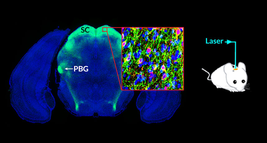
BUNDLE OF NERVES  When parvalbumin-positive neurons (pink in box) nestled in the mouse brain’s superior colliculus (SC) were stimulated by a laser, they carried threatening information to a brain region called the parabigeminal nucleus (PBG). Image shows a slice of the brain from the top of the head to the bottom. 

C. Shang et al/Science 2015

View the video

A zap of blue light to the brain can make a mouse flee or freeze. By exciting select nerve cells, scientists have for the first time identified and hijacked a neural pathway that carries threatening information from the eyes to the brain.

The results, published in the June 26 Science, may help scientists better understand how the brain handles fear. That understanding may ultimately lead to ways to help people with abnormal responses to fear, such as those with post-traumatic stress disorder, says study coauthor Peng Cao of the Chinese Academy of Sciences in Beijing.

Cao and colleagues studied nerve cells in the superior colliculus, a waystation between the eye’s retina and more complex areas of the brain. The team engineered select groups of these nerve cells to fire off signals in response to blue light, delivered by slender optical fibers implanted in the brain.

When the light stimulated a certain type of nerve cell, called parvalbumin-positive neurons, the animals ran around frantically as they attempted to escape. Then the mice held perfectly still for nearly a minute, even after the light was turned off. Those reactions suggest that the stimulated neurons carry fear signals.

These neurons then send threat signals to a different brain region called the parabigeminal nucleus, the researchers found. The signal then moves to the amygdala, an almond-shaped bundle of neurons already known to be involved in detecting threats.

Male mice zapped by brighter and longer light spent more time trying to escape and less time freezing than males exposed to less light. “This is generally consistent with the idea that animals tend to escape rather than fight when the threat is extremely strong,” Cao says.

In general, females seemed more prone than males to escape rather than freeze, the team found. Because the neural pathways appeared to be the same in males and females, the researchers suggest that other brain regions or hormones may be behind the different reactions.

Illuminating this neural pathway has revealed part of the link between the visual scenes in the outside world and the brain centers that trigger fear responses, says neuroscientist Simon Hippenmeyer of the Institute of Science and Technology Austria in Klosterneuburg. Further clarifying this neural pathway may lead to a deeper understanding of how the brain translates danger into action, he says.

Neurons in the superior colliculus seemed to be responding to a particular sort of scary signal — an approaching object. Separate experiments on anesthetized mice revealed that these neurons responded as a virtual soccer ball rolled toward the animal. (Although the mice were knocked out, one eye was propped open and could still respond to visual input.) Similar neurons have been found in locusts, fish and fruit flies, says neuroscientist Fabrizio Gabbiani of Baylor College of Medicine in Houston.

Detecting and responding to an approaching object is a very basic and important feature of brains. In humans, it helps people cross the street or avoid a car crash, Gabbiani says. “This is one of the first things that the visual system was designed to do.”

FROZEN WITH FEAR  After blue light stimulates nerve cells in the brain’s superior colliculus, a male mouse tries to escape a faux threat and then freezes for about a minute. Credit: C. Shang et al/Science 2015

Laura Sanders is the neuroscience writer. She holds a Ph.D. in molecular biology from the University of Southern California.

More Stories from Science News on Neuroscience

From the Nature Index

Paid Content