In Pixels and in Health
Computer modeling pushes the threshold of medical research
Moment by moment, a movie captures the action as a group of immune cells scrambles to counter an invasion of tuberculosis bacteria. Rushing to the site of infected lung tissue, the cells build a complex sphere of active immune cells, dead immune cells, lung tissue, and trapped bacteria. Remarkably, no lung tissue or bacterium was harmed in the making of this film.


Instead, each immune cell is a computer simulation, programmed to fight virtual tuberculosis bacteria on a square of simulated lung tissue. In their computer-generated environment, these warrior cells spontaneously build a structure similar to the granulomas that medical researchers have noted in human lungs fighting tuberculosis.
The simulation, created by Denise Kirschner of the University of Michigan in Ann Arbor, is an example of an emerging technique called agent-based modeling. This new tool in the world of medical research relies on computing power instead of tissues and test tubes. A growing cadre of researchers, including Kirschner, predicts that agent-based modeling will usher in a broadened understanding of complex interactions within the human body.
The agents in the models are individual players—immune cells in the tuberculosis example. Each player is programmed with rules that govern its behavior. Computer-savvy researchers then set the agents free to cooperate with, compete with, or kill each other. Meanwhile, the agents must navigate the surrounding environment, whose properties can vary over space and time.
Scientists can manipulate disease progression within the models by changing the agents or their environment and then watching what happens. As opposed to traditional, biologically based in vivo or in vitro experiments, these computer trials are dubbed “in silico.” The results can suggest biological experiments to test the models’ findings and may eventually lead to new medical treatments.
Even simple rules assigned to agents can give rise to surprisingly complex behaviors. When many independent agents interact, they create phenomena—such as the granulomas—that can’t necessarily be predicted by breaking down the system into its separate components, says complex-systems specialist John Holland of the University of Michigan.
You’ve got to study the interactions as well as the parts,” Holland says.
In-silico modeling differs from traditional mathematical modeling, which uses differential equations to understand how molecules or cells behave in an averaged, continuous way. Instead, the agents of in-silico modeling make independent decisions in response to situations that they encounter. As a result, unusual activity of even a small number of cells can change the entire system’s behavior.
Computers can now calculate thousands of interactions with ease, says Alan Perelson of Los Alamos National Laboratory in New Mexico. “Agent-based modeling has only come into its own with the arrival of really powerful computers sitting on people’s desktops, within the last 10 or 15 years,” he notes.
Pioneered for economics and population-dynamics studies (SN: 11/23/96, p. 332: http://www.sciencenews.org/pages/sn_arch/11_23_96/bob1.htm; and Science News Online: “Simulating Society” at http://www.sciencenews.org/pages/sn_arc99/4_10_99/mathland.htm), agent-based modeling has only recently plumbed the inner workings of the human body, Perelson adds. That’s partly because new imaging and genetic techniques are providing crucial data on which agents’ rules can be based.
“Agent-based modeling represents a new frontier with respect to how we do science,” says surgeon Gary An of Cook County Hospital in Chicago. “In medicine in particular, all the diseases that we’re now dealing with are complex problems: sepsis, cancer, AIDS. All these things are disorders of the system as a whole.”
Inflammation simulation
An, whom Kirschner calls an in-silico “groundbreaker,” got into agent-based modeling to help people survive traumatic injuries and major infections.
A leading cause of death for patients in intensive care units, An explains, is a syndrome called systemic inflammatory response syndrome/multiple organ failure (SIRS/MOF), also termed sepsis when it occurs in response to an infection. In this syndrome, the body’s inflammatory response rages out of control after a severe injury or bacterial infection. Excessive inflammation can kill a patient by attacking and shutting down vital organs. More commonly, the runaway inflammation paralyzes the rest of the immune response, and the patient then dies of secondary infections.
During the 1990s, researchers performed clinical experiments in an attempt to develop drugs that dampen an overwhelming inflammatory response to injury, An notes. Only one drug, activated protein C, appeared to help patients with SIRS/MOF. An suggests that trials of other drugs failed because they were planned using data representing individual components of the inflammatory response rather than the interactions of the immune system as a whole.
An says, “It’s kind of a Humpty Dumpty syndrome, where after you break the system apart, you can’t put it back together.”
An turned to agent-based experiments to understand how the body’s inflammatory processes work together to generate SIRS/MOF. In a seminal paper in 2001, he described a model of the inflammatory response that included all the information that he could find about the immune system and inflammation.
By assigning inflammation-implicated cells as agents in an environment that simulated the body’s circulatory system, An reproduced the four typical trajectories of SIRS/MOF. At a low concentration of bacteria, the inflammatory response killed the infection and the virtual patient recovered. High concentrations of infectious bacteria overwhelmed the simulated system, and it died.
At moderate concentrations of bacteria, the model replicated the two trajectories of SIRS/MOF of most interest to medical researchers: organ failure and immune paralysis due to excessive inflammation. Although An says that his model is “very much in its infancy,” he and his colleagues have used it to simulate the trials of some of the potential SIRS/MOF drugs that were conducted in the 1990s.
Employing only the biological data available when the trials were designed, the agent-based model could have predicted the trials’ failures, the team reported in 2004. “It’s not to say you necessarily would not have done the trials,” An notes. “But if you had done [the modeling] before you got to the clinical trials, you might have gone back and relooked at some of your assumptions.”
Laptop laboratory
The new-style models contain four components: the agents, their rules, the agents’ environment, and the time scale on which they operate. The environment is usually represented by a grid in which each square is programmed to contain data, such as a concentration of molecules or virus particles. The agents themselves are shown as colored dots that can migrate from square to square on the grid.
For instance, in Kirschner’s simulation of tuberculosis infection, immune cells are the agents, and the grid represents a 2-millimeter-square patch of lung tissue, big enough to hold a nascent granuloma. Each of 1,000 squares on the grid contains chemical and structural information about the tissue as well as some concentration of the tuberculosis pathogen, Mycobacterium tuberculosis.
“You can give structure and character and rules to that lung tissue so that if a[n immune] cell is in a particular spot, it has to behave in a certain way,” Kirschner explains.
Bright dots scurry over the grid, representing immune cells called macrophages and T cells. Macrophages capture and sometimes destroy foreign particles such as dust or bacteria. T cells, meanwhile, communicate with the macrophages to make them more aggressive and marshal additional immune efforts.
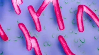
Worldwide, tuberculosis is the infectious disease that causes the most deaths, 2 million to 3 million per year. An estimated one-third of the world’s population is infected with the pathogen. The bacteria tend to hide out inside macrophages. Unless T cells activate the macrophages to destroy their stowaways, the bacteria multiply, eventually causing the macrophages to burst and release their bacterial load to other cells.
Once infected with the pathogen, about 5 percent of infected people come down with acute tuberculosis right away. Most people, however, develop a latent form of the disease. Of these, only 10 percent eventually develop full-blown tuberculosis.
The reasons for people’s different responses to tuberculosis infection remain a mystery, though all infected people form granulomas in their lungs, says Kirschner. Scientists have studied granulomas in various stages of formation, but they’ve never witnessed the process that creates them.
Granulomas in patients with acute tuberculosis expand and build up large proportions of dead, or necrotic, tissue in their cores. Using their agent-based model, Kirschner and her colleagues found that out of 27 parameters examined, only 7 strongly affected whether a granuloma would turn necrotic. The team described its model and results most recently in the May 2005 Trends in Microbiology.
For example, the timing of T cell arrival to infected lung tissue partly controlled its fate. Immediate arrival cleared the infection completely and prevented a granuloma from forming, while delayed arrival produced necrotic granulomas.
At moderate T cell–arrival times typical of normal human lungs, most of the virtual patients developed granulomas that contained the infection and had little necrotic tissue. However, the granulomas turned necrotic in about 5 percent of the trials—the same percentage of the human population that immediately develops the acute disease. The results suggest that patients would benefit from therapies that encourage rapid recruitment of T cells to sick lungs.
Experimental biologist Victor DiRita of the University of Michigan says that Kirschner’s approach to tuberculosis will aid experimental efforts to understand the disease. “You can’t scoff at the fact that they’ve now been able to grow a granuloma in a computer that has a lot of the structural characteristics, as far as we can tell, of a real granuloma,” DiRita comments.
Cancer conundrum
An extreme malfunction of human cells occurs during cancer. A tumor arises when a normal cell mutates into a cancer cell, which replicates uncontrollably. Tumor cells can eventually migrate to other parts of the body.
Because cancer cells interact in complex ways with their environment, with other cancer cells, and with normal cells, they make a perfect target for agent-based modeling, says Thomas Deisboeck of the Massachusetts Institute of Technology.
Deisboeck and his colleagues apply agent-based modeling to the behavior of brain-tumor cells. These and other cancer cells tend either to proliferate, causing a tumor to grow, or to migrate to a new location, but they seldom do both at once. The reasons behind a cell’s behavioral choice aren’t known.
Deisboeck’s team examined cell responses to a molecule called epidermal-growth factor (EGF), which reaches high concentrations in tumors. EGF influences cell proliferation and migration, but no study had looked at the molecule’s effect on both behaviors simultaneously. Deisboeck’s group simulated cancer cells as agents, subjecting them to fluctuating concentrations of EGF and associated molecules that affect EGF’s behavior.
In the model, these molecules controlled when cancer cells switch between proliferation and migration. Moreover, high densities of receptor sites for EGF on cell surfaces made simulated tumors expand faster. The team reported its results in the April 21, 2005 and the Jan. 7, 2006 Journal of Theoretical Biology.
Cancer researcher Ken Pienta of the University of Michigan praises Deisboeck’s work. “He’s able to explain cellular actions based on simple molecular rules. That gives you a perspective that’s going to be critical for therapeutic development.”
Pienta, in collaboration with Holland, has also created agent-based models of cancer. They cast mutations, instead of cancer cells, as agents. Depending on how mutations interact within a given cancer cell, the cell may or may not survive and propagate.
Although Pienta and Holland haven’t published their agent-based efforts, the simulation has influenced Pienta’s thinking as he designs biological experiments. He says, “What the modeling does is it forces you to push your preconceptions out the door.”
Digital patients
Pienta notes that in-silico modeling by itself won’t provide clinical advances. Biological experiments, both in vitro and in vivo, remain crucial for developing therapies for disease.
Agent-based modeling can suggest possible experiments, predict which hypotheses are most likely to be true, and integrate data provided by experimental biologists, says Deisboeck. Most modelers collaborate with experimentalists, who provide biological data and embark on flesh-and-bone trials to test the models’ findings.
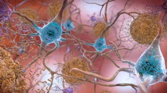
“The idea is to validate your results against experimental data,” says Leah Edelstein–Keshet of the University of British Columbia in Vancouver, who uses agent-based modeling to simulate Alzheimer’s disease. She’s now collaborating with drug company Merck to pursue potential therapies.
“You can imagine a future where you could interact with the agent-based models like you can in a video game,” speculates Edelstein–Keshet. For instance, and use the results to decide treatment for the real patient. But Edelstein-Keshet and others caution that such a future remains a long way off.
“The real-life systems we’re trying to understand are just immensely complex,” says biostatistician Thomas Kepler of Duke University in Durham, N.C. “How things behave inside the body is very hard to predict based on experiments done outside the body.”
However, many scientists expect in-silico modeling to play an ever-larger role in medical research. As agent-based models are combined with more-traditional mathematical descriptions of disease processes, their predictive power will grow.
“I have a strong belief that agent-based modeling is going to be a very powerful tool to analyze biology,” says Kirschner. “You’re really allowed to give the objects in your programming a life of their own.”