X Ray Excels: Technique brings a new image to medicine
Imaging soft tissue in detail has required enormous particle accelerators that span several city blocks. A new method may soon bring this valuable diagnostic capability into hospital settings. There, researchers say, it will provide physicians with unprecedented power to spot tumors, clogged arteries, and other soft-tissue problems.
Researchers have long known that phase-contrast X-ray imaging can yield stunning pictures of soft tissue. Currently, however, the technique requires X rays from a synchrotron, a particle accelerator measuring 300 meters or more in diameter.
“Phase-sensitive X-ray imaging can produce images that show much more detail in tissue structure, but none of the techniques could be used efficiently in a hospital-based setup with small X-ray sources,” says Franz Pfeiffer of the Paul Scherrer Institute in Villigen, Switzerland.
Now, his team reports an advance that it says will enable radiologists to perform phase-contrast imaging using conventional hospital X-ray machines. The team describes its innovation in the Aug. 8 Optics Express, an online journal.
“The beauty of this is … it could potentially allow you to use more-normal sources of X rays, like X-ray tubes,” comments L. Dean Chapman of the University of Saskatchewan in Saskatoon.
Conventional X-ray techniques take advantage of the fact that materials in the body absorb X rays to different extents. Bone, for instance, absorbs more X rays than soft tissue does.
However, different types of soft tissue, such as a cancerous patch of breast tissue and a normal patch, often have only tiny, hard-to-detect variations in absorption, says Pfeiffer. As a result, soft-tissue imaging methods such as mammography must use high doses of radiation, which can pose health risks for the patient, to yield even low-resolution images.
Phase-contrast X-ray imaging relies instead on refraction, or changes in the angular trajectory of X rays. Just as light rays bend when they enter water from air, X rays deflect as they travel through objects of varying densities. This deflection can be precisely measured to produce highly detailed images.
Chapman notes that phase-contrast imaging can use lower-wavelength X rays than conventional methods do. Because the low-wavelength rays tend to pass through tissue, the procedure delivers a much lower radiation dose to the patient than do standard X-ray methods.
“The problem is, it’s hard to do,” Chapman says.
Pfeiffer says that his team has simplified the task with a better analyzer for refracted X rays. The team uses a pair of gratings, each scored with 2-micron-wide furrows, to produce an interference pattern from deflected rays. A detector then produces an image from this pattern. Team member Christian David invented the grating.
Previous analyzers could resolve only a very intense X-ray beam, a beam of a single wavelength, or a beam of highly parallel rays. Coauthor Timm Weitkamp of the European Synchrotron Radiation Facility in Grenoble, France, says the new gratings can handle the less intense, multiwavelength, and multidirectional beams that emerge from typical hospital X-ray tubes.
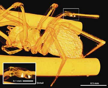
As a demonstration, Weitkamp and his coworkers have used their grating with a synchrotron source to image the leg joints of a small spider. They say that the first experiments using a standard X-ray tube indicate that their technique also could be used in clinical settings.