Brain discoveries open doors to new treatments
 For centuries, scientists have strived to figure out the workings of the human brain, but that blob of matter tucked inside a bony shell long resisted efforts to divine its secrets.
Techniques invented in the early 1900s, including angiography and electroencephalography, made it possible to examine some characteristics of the brain without invading the skull. But it wasn’t until the 1970s, with the development of functional PET and MRI scanning, that it became possible to see the brain in action.
Two stories in this issue illuminate just how far we’ve come in being able to explore and influence the brain in this extraordinary era of neuroscience innovation. “Brain-zapping implants that fight depression are inching closer to reality,” by neuroscience writer Laura Sanders, examines efforts to treat depression with electrical stimulation. Rather than deliver a whole-brain zap, which is used in electroconvulsive therapy for severe depression, these experiments are testing whether nudging parts of the brain with electrodes could work better than current treatments.
“It’s one of those things that sounds a little bit like science fiction, but they’re actually doing it,” Sanders told me. “What if we could have this really precise, gentler, kinder approach that worked?”
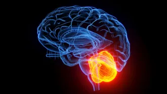
A second story by Sanders looks at recent discoveries about the cerebellum, a brain structure that had been thought to be involved only in movement. Now scientists say that the cerebellum plays a role in many of the things that make us human, including memory, language and social relationships.
Improved technology, such as the tiny electrodes used in the brain-zapping therapy, drives most of these discoveries. And often the innovation comes from combining methods.
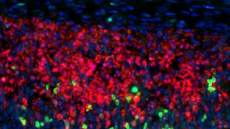
In January, Sanders wrote about researchers affiliated with Howard Hughes Medical Institute’s Janelia Research Campus who managed to zoom in on the structure of just one nerve cell at a time by combining a laser lattice light-sheet microscope with a technique called expansion microscopy, which enlarges tiny tissue samples to reveal individual structures. And a group of Stanford University scientists created an advanced form of optogenetics that uses laser light to control individual nerve cells. The researchers used the technique to switch nerve cells on and off in mouse brains, controlling the animals’ behavior (SN Online: 1/17/19).
Sanders appreciates the difficulty of these achievements. She has a Ph.D. in molecular biology, which she earned by spending many, many hours tweaking nerve cells in fruit fly brains to see how various regions are involved in the flies’ mating dance. “But I got really tired of watching videos of fruit flies mating,” she says. Plus doing all those dissections of tiny fruit fly brains meant she needed glasses by the time she finished her degree.
Ultimately, Sanders realized, she’s much more interested in the brains of people, and she decided to become a science journalist. “That’s part of what I love,” she says. “Thinking about how complicated human behavior is, and how we’re actually getting clues now that were previously impossible.” We’re glad she decided to use her deep understanding of neuroscience to explain it to the public. The fruit flies’ loss is our gain.