New ways to image and control nerve cells could unlock brain mysteries
Using the methods to target neurons in mice and fruit fly brains may lead to insights
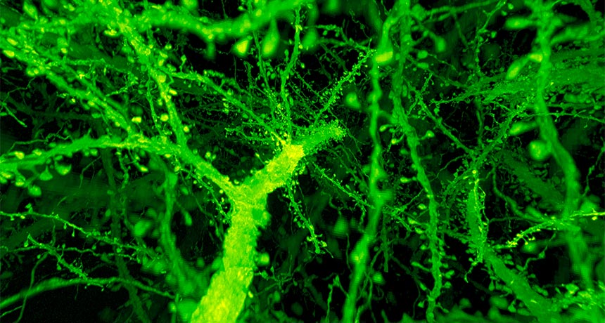
TINY REVEAL A powerful new technique reveals tiny, message-receiving bumps that dot nerve cells’ surfaces (green), called dendritic spines, in a mouse brain.
R. Gao et al/Science 2019
Using laser light, ballooning tissue and innovative genetic tricks, scientists are starting to force brains to give up their secrets.
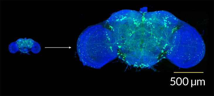
By mixing and matching powerful advances in microscopy and cell biology, researchers have imaged intricate details of individual nerve cells in fruit flies and mice, and even controlled small groups of nerve cells in living mice.
The techniques, published in two new studies, represent big steps forward for understanding how the brain operates, says molecular neuroscientist Hongkui Zeng of the Allen Institute for Brain Science in Seattle.
“Without this kind of technology, we were only able to look at the soup level,” in which diverse nerve cells, or neurons, are grouped and analyzed together, she says. But the new studies show that nerve cells can be studied individually. That zoomed-in approach will begin to uncover the tremendous diversity that’s known to exist among cells, says Zeng, who was not involved in the research.
“That is where the field is going. It’s very exciting to see that technologies are now enabling us to do that,” she says.
These novel abilities came from multiple tools. At Howard Hughes Medical Institute’s Janelia Research Campus in Ashburn, Va., physicist Eric Betzig and his colleagues had developed a powerful microscope that can quickly peer deep into layers of brain tissue. Called a lattice light sheet microscope, the rig sweeps a thin sheet of laser light down through the brain, revealing cells’ structures. But like any microscope, it hits a wall when structures get really small, unable to resolve the most minute aspects of the scene.
A trick that expands the tissue like a balloon under the microscope solved that problem. The method, called expansion microscopy, makes tiny samples easier to see by infusing them with a gel that swells, says neuroscientist Edward Boyden, an HHMI investigator at MIT whose lab developed the technique. The gel preserves the tissue’s structure while also expanding it.
Turning the powerful lattice light sheet microscope on expanded fruit fly brains and sections of mice brains revealed features of individual nerve cells, the researchers report in the Jan. 18 Science. The team counted cell connections called synapses, saw how a fatty substance called myelin wrapped around nerve cells’ message-sending extensions and pinpointed all the nerve cells that produce the chemical messenger dopamine.
Those meticulous views will allow more experiments, says Betzig, now an HHMI investigator at the University of California, Berkeley, such as studying whether synapses look different in certain diseases, or how myelin forms during development. “If you want to dive deep into any one of these areas, you can do it now,” he says.
Along with these new details on nerve cell anatomy come hints about some of these cells’ jobs. Karl Deisseroth, a psychiatrist and neuroscientist at Stanford University, and colleagues developed an advanced form of optogenetics, a technique that uses laser light to control genetically engineered nerve cells. With advances in microscopy and improvements to a protein that responds to laser stimulation, the researchers were able to monitor individual nerve cells’ behavior and activate them at will, changing the mice’s eating behavior. The results, described online January 16 in Nature, help to untangle cells involved in eating behavior and social experiences.
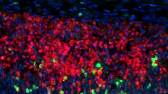
In the study, the researchers targeted nerve cells in mice’s orbitofrontal cortex, a stretch of tissue on the outer front surface of the brain. Because the cells involved in eating behavior and in social behavior are mixed together there, they’re not easy to study separately.
So Deisseroth’s team used genetic tricks to identify single nerve cells that are active as a mouse does a certain behavior — in this case, licking high-calorie water or interacting with another mouse. After identifying certain cells, the researchers then used laser light to prod the cells into action and watched for the resulting behavior. When the scientists stimulated a handful of the “eating” nerve cells, mice licked up more of the calorie-dense water. But when the team stimulated the social nerve cells, licking decreased, results that hint that social interactions can curb eating behavior.
Deisseroth and his colleagues first described the method for stimulating single nerve cells in 2012, but until now, hadn’t been able to use it to control behavior in a mammal. Advances in microscopy, including a special lens that sits atop the brain and focuses light in a particular way, allowed the researchers to stimulate nerve cells about three millimeters deep in the brain of a live mouse — the deepest single cell stimulation to date, the researchers report.
“Most of the mouse brain is deep and difficult to probe,” Deisseroth says. The new method “opens the door to versatile investigation of the entire mammalian brain,” he says.