Busy neurons don’t always draw blood

Mouse study suggests caution in interpreting functional MRI results

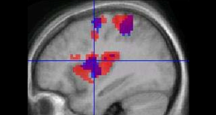
GO WITH THE FLOW  Functional MRI scans use blood flow (colors represent changes in blood flow) to indicate nerve cell behavior, but those two things aren’t always coupled, a new study finds. 

K. Roberts et al/BMC Anesthesiol. 2008/Open-I (CC BY 2.0) 

A mainstay of many neuroscience labs, functional MRI relies on blood flow changes in the brain to serve as proxies for active nerve cells. But a new study on mice finds that neurons can be busy with no hint of blood-flow changes.

Many researchers assume that fMRI signals reflect neural activity, says study coauthor Patrick Drew, a neuroscientist at Penn State, “that when neural activity goes up, you should see increases in blood flow.” But recently, that cozy relationship has come under increasing scrutiny.

The results, published in the Aug. 13 Journal of Neuroscience, emphasize the need for caution when interpreting brain-scan results, says neuroscientist Shella Keilholz of Georgia Tech and Emory University School of Medicine in Atlanta.

Functional MRI detects tiny changes in the brain’s amount of oxygenated blood, which researchers often interpret as signs of neurons sending off more electrical messages.  And in some cases, that interpretation is correct. But by finding a case when neurons are busy with no corresponding change in blood movement, the new study shows that blood flow isn’t always a reliable marker of neural activity. “The picture is getting more and more complicated,” Keilholz says.

To tease apart the individual actions of blood flow and neuron firing, Drew and colleagues separately measured each process as mice voluntarily walked on a treadmill. Brain activity in a region called the somatosensory cortex, which is involved in sensing the environment, behaved as expected: Neurons became active while the animals walked, electrodes revealed. And the somatosensory cortex experienced a corresponding surge of blood, experiments on other mice showed. 

But in a different part of the brain, the relationship between neuron activity and blood flow disappeared. Neurons in the frontal cortex, a region involved in complex thinking as well as certain aspects of movement, were active while the mice walked. But blood flow didn’t budge, Drew and his colleagues found.

The decoupling shouldn’t come as a shock, says neuroscientist Elizabeth Hillman of Columbia University, who has also found evidence that neural behavior isn’t always yoked to blood flow. One study by other researchers on awake monkeys found that blood flow can increase with no corresponding neural activity, although that result remains controversial. Other evidence comes from experiments on anesthetized animals, not on awake brains.

Scientists don’t understand all of the various signals that control blood flow in the brain. Experiments on neurons in a dish have found that the behavior of certain kinds of neurons can cause blood vessels to dilate while other neurons’ activity causes vessels to constrict, Drew says. Blood flow is probably influenced by complex mixtures of signals that change depending on brain region. 

Editor’s note: This story was updated on August 22, 2014, to note that the mice walked, not ran, on the treadmill.

More Stories from Science News on Neuroscience

From the Nature Index

Paid Content