Cataloging the connections
Viewing the brain as a network may help scientists tackle its complexity
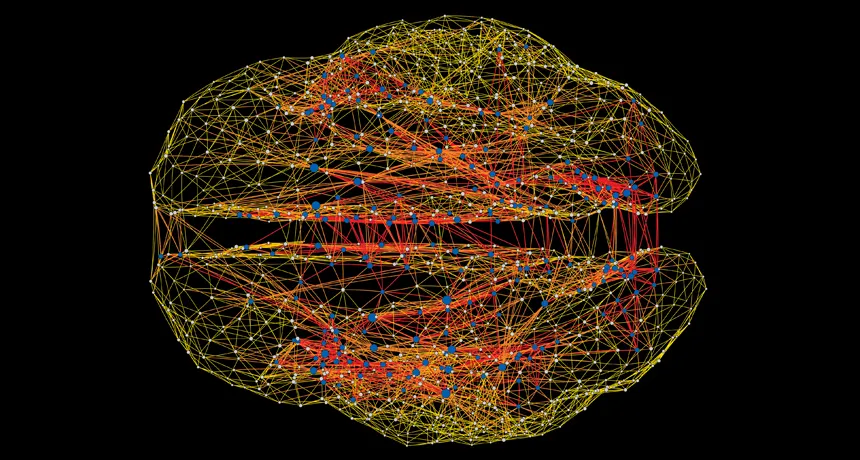
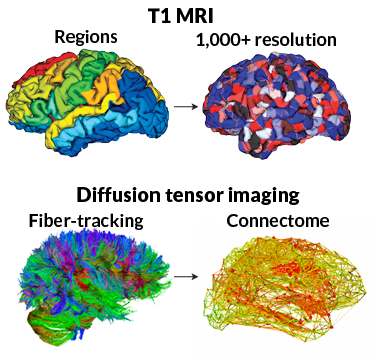
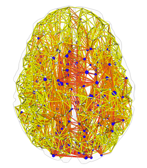
LINKED IN Using brain scanning technologies, scientists can create maps (above) showing the brain’s wiring, consisting of white matter fibers that link different parcels of the brain’s gray matter. The most highly connected parcels, or hubs, are indicated by blue and white dots.
Courtesy of M.P. van den Heuvel
Mapping the human brain is a noble goal, but a rather ill-defined one. It’s like making a map of the United States. You could just show political boundaries and the locations of cities. Or you might depict geographical features like mountains and rivers. Or transportation routes, like interstate highways and railroad tracks. You might even go Google Maps all the way and show the location of every individual house.
The brain possesses a similar diversity of scale: two hemispheres of convoluted gray matter, each with four regional lobes, traversed by superhighways of white matter fibers communicating with billions of individual cells. So some brain maps focus on outlining anatomical areas, others track the white matter wiring, still others divide the gray matter into tiny parcels and record their activity during different mental tasks. But eventually, scientists want to map everything. Their ultimate goal is a catalog of all the connections between all the brain’s cells and regions, a master map known as the connectome.
It’s a formidable task, comparable to identifying every building in the country and then tracing the routes of all the people and cars that travel among them. Yet mapping all those connections promises a huge potential payoff, many researchers say, and it will be essential to pursuing the even grander goals articulated by President Obama for understanding how the brain thinks and learns (SN: 5/4/13, p. 22).
“The BRAIN Initiative of President Obama emphasizes determining connectivity,” says neuroscientist Scott Emmons of Albert Einstein College of Medicine in New York. “And clearly we won’t be able to understand the nervous system unless we know this connectivity.”Stanford University neuroscientist William Newsome, cochair of the National Institutes of Health panel establishing priorities for the president’s project, agrees.
“This is what we interpreted the overarching goal of the BRAIN Initiative to be,” says Newsome, “to map the circuits of the brain, measure the fluctuating patterns of electrical and chemical activity flowing within those circuits and to understand how their interplay creates our unique cognitive and behavioral capabilities.”
Tough as that challenge seems, substantial progress has already been made. Scientists have completely described the connections in the primitive brain of the tiny roundworm Caenorhabditis elegans, for instance. Human studies have begun to map the white matter fibers that physically link various brain regions. Brain scans are revealing which regions operate in synchrony, a further clue to how they are connected.
And using the mathematical theory describing networks, scientists have begun to perceive how brain cells cooperate to generate thought and behavior. In fact, network math suggests that deep insights into the brain’s connections are possible even without mapping all the links for every single cell.
Ultimately such insights should assist in diagnosing and treating a number of brain diseases, such as schizophrenia, that result from faulty connections. “It’s been suggested,” says Emmons, “that some severe disorders such as schizophrenia and autism are in fact connectopathies.”
A simple brain
In principle, the human connectome consists of literally every single link between every single nerve cell, or neuron, in the brain. But such a complete map is technologically out of reach at the moment. With a neuron population of roughly 85 billion, each maintaining thousands of connections, the connectome comprises an unfathomably vast network, with hundreds of trillions of links. So in humans, connectome research focuses on links between anatomical brain regions or just small parcels of brain tissue. The Human Connectome Project, launched by the National Institutes of Health in 2010, maps portions of gray matter on the cubic millimeter scale, roughly the size of a grain of salt. It’s like mapping roads connecting cities and towns but ignoring side streets and individual houses.
An additional wrinkle of complexity distinguishes between physical links via white matter (cellular projections sheathed in myelin that wire regions together) and functional connections, identified by which brain regions are simultaneously active when performing a specific task.
This “functional connectome” is closely related to physical links, of course. But the human brain’s complexity makes it infeasible to track that relationship on the scale of individual neurons. So scientists have sought some simpler substitutes to get insights into how neurons interact.
A favorite for this purpose is Caenorhabditis elegans, which possesses one of the simplest brains in the biosphere. A male C. elegans possesses 383 neurons (the hermaphrodite has even fewer), allowing scientists to catalog all the worm’s neurons and trace their more than 2,000 connections using electron microscopy.
Worms and people share common ancestors, suggesting that the worm can provide information about how the human brain evolved. Although separated by eons of evolution, worms do use some of the same cellular messenger chemicals and display other properties recognizable in humans, Emmons pointed out in November at the annual meeting of the Society for Neuroscience. Both worm and human brain can be described, for example, by the mathematical theory of networks, officially known as graph theory. In graph theory, networks are represented by dots connected with lines; the lines are called links (or edges) and the dots are called nodes (or vertices). In C. elegans, the nodes are neurons, linked by the synapses through which the neurons communicate. Network math allows calculation of various properties, such as how many links a neuron makes on average to other neurons, and the minimum number of connections needed to transmit a signal from one neuron to any other one in the network.
Network analyses of C. elegans have revealed how sets of highly interconnected neurons can function as a module to govern a behavior such as mating. Using network math to discern the brain’s modular organization might work in people as well as in worms, suggesting that connectome research may be useful even without mapping all the synapses, Emmons noted.
“Do we have to identify every single synapse to understand brain connectivity, or can we find computational subunits of neurons that do similar functions and treat them as a group?” he said at the neuroscience meeting. It may be possible, he believes, to show how networks are built from groups of neurons without the need to determine all the wiring within each group. “This is certainly a challenging and exciting goal as brain connectomics goes forward,” Emmons said.
Neural hubs
One important finding is that worm brain connectomes are “small world networks,” famous for how few steps it takes to link any two nodes (SN: 2/17/07, p. 104). Such networks typically possess highly connected nodes, or hubs, that help shuffle signals from one node to another efficiently.
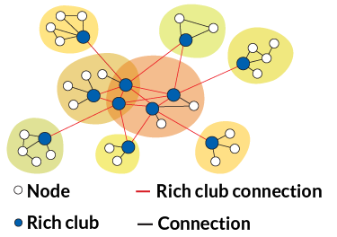
In the C. elegans hermaphrodite, 11 neurons have been identified as “rich club” hubs, neuroscientist Edward Bullmore of the University of Cambridge and collaborators reported last April in the Journal of Neuroscience. These hubs are not only well-connected in their own network, but are also connected to each other, forming a network, or club, of highly connected nodes.
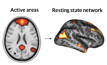
Rich club nodes also exist in the human brain, even though the nodes are parcels of brain tissue rather than individual neurons. Similarly to social networks like Twitter, the brain’s network consists of “communities” of anatomical regions that share information and participate in common tasks. Brain scans have identified several such communities, called resting state networks, that are related to important brain functions, such as vision, movement, hearing and touch. But the various resting state networks are not tightly connected to each other. So the brain needs a system to coordinate its various tasks and transmit information from region to region. That’s where the rich club hubs enter the game.
In humans, rich club hubs are found in various brain regions, no matter their job. “Hubs tend to be present in all functional domains of the brain,” says Martijn van den Heuvel of University Medical Center Utrecht in the Netherlands.
He and Olaf Sporns of Indiana University investigated the rich club hubs by mapping the white matter in 75 people. Out of 1,170 parcels of gray matter in each brain, on average 17 percent were highly connected enough to be considered rich club hubs. Such hubs were found in all 11 of the resting state networks examined in the study. Often those richly linked hubs showed up in “confluence zones” where resting state networks overlapped on the brain’s surface layer.
Not only are the rich nodes highly connected within their network, they are also highly connected to each other — which is what makes them members of a club, van den Heuvel and Sporns found. “What we observe is that the level of connectivity between those hubs was around 40 percent higher than what we would expect,” van den Heuvel said at the neuroscience meeting.
Their presence in all functional networks, along with their club membership, indicates that these rich club hubs anchor the brain’s data sharing system, merging information from the various functional networks.
“In the brain or in neural systems, communication is not just sending a UPS package around with the content of the package remaining the same,” van den Heuvel said. Data from different regions are always being combined and coordinated.
Understanding the rich club will aid efforts to comprehend such high-level brain processes, van den Heuvel believes. Thought and consciousness, for instance, may involve a “global workspace” in the brain that relies on the network of rich club connections to manage communication among the brain’s many regions.
“The global workspace does not coincide with a single anatomical or functional module in the brain, but rather involves a widely distributed neural system of long-distance anatomical projections,” van den Heuvel and Sporns wrote last September in the Journal of Neuroscience.
Brain as network
Already the network approach to understanding brain connections has led to insights about important mental functions such as learning and memory. One relevant finding is that the brain’s networks are not static, but capable of rapid changes over time. And some networks are more flexible than others.
“Within the brains there are some regions that tend to be very rigid,” says Danielle Bassett of University of Pennsylvania. “Core” regions involved in sensations and motions are typically rigid, she says, while peripheral systems, involved in thinking and making visual associations, tend to be more flexible.
“People who have a more rigid core and more flexible periphery are those who learn better than individuals whose core is more flexible or whose periphery is more rigid,” says Bassett. “So this separation of functions between core and periphery is actually quite important for how individuals learn.”
Besides illuminating the workings of the normal brain, studies of network structure and function have also led to insights into brain disorders. Van den Heuvel points out that the central role of the rich club makes it a prime suspect in cases when the brain goes awry.
“If it’s such a central system, one would suspect that abnormal wiring of the system might lead to brain dysfunction, and we indeed have some evidence that it is happening in schizophrenia and Alzheimer’s,” he says.
Some of that evidence comes from Bassett and collaborators. They have found numerous differences when comparing the brain networks of healthy individuals with those of schizophrenia patients. At the neuroscience meeting, Bassett outlined several findings from various groups over recent years. One major difference is in the strength of connections, measured by how likely linked nodes are to be simultaneously active, in schizophrenia versus healthy brains.
“Across practically every single area of the brain we see a decrease in strength in the schizophrenia networks as compared to those of controls. That suggests that there can be a very global decrease in communication constructs in schizophrenia,” she said.
On the other hand, while connections are generally weaker, they are also more variable. In a healthy brain, highly connected hubs tend to have strong connections, less connected nodes have weaker connections. But in schizophrenia, a given hub will have some strong connections but also some weak ones, suggesting a lack of proper brain organization.
“Potentially we could interpret this as healthy controls are able to separate the functions that different brain regions have to perform well, while schizophrenics are not able to segregate those functions in the same way because their networks are disorganized,” Bassett said.
Furthermore, it is in the brain’s weaker connections where schizophrenia patients differ most from healthy people. Patterns of weak connections distinguish schizophrenic brains from healthy ones with 75 percent accuracy, Bassett and colleagues reported in 2012 in NeuroImage. And the weak connections differ in terms of their anatomical locations, often touching areas of the brain known to be involved in schizophrenia’s symptoms. Measures of schizophrenia symptoms involving attention, memory and negative affect were all strongly related to networks of weak connections.
Similar network-based studies have begun to identify important features of ADHD and autism, other brain disorders believed to be related to faulty connections. In ADHD, connections are sparser than normal and medications appear to work by repairing the brain’s network structure, Damien Fair, of Oregon Health & Science University in Portland, reported at the neuroscience meeting. In autism, on the other hand, nodes appear to be more connected than usual.
Understanding the connectome in all its complexity may not lead to immediate cures for brain diseases. But it’s becoming clear that progress in fighting brain disorders, and understanding the normal brain, will not be possible without embracing graph theory — the mathematics of networks — for analyzing the brain’s connections.
“In general the brain is indeed a network and we should approach it as such,” says van den Heuvel. “And graph theory may be one of those techniques or tools to extract properties that might provide more information on how brain function can emerge from the underlying anatomy.”
