Coming to Terms with Death

Accurate descriptions of a cell's demise may offer clues to diseases and treatments

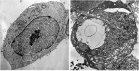
Death is a part of living�and an essential one. From conception onward, cells divide over and over again. Their endless proliferation would quickly lead to elephantine bodies were it not for a compensating death of cells.

Thriving cancer cell (left) lacks the vacuoles–clear, fluid-filled patches–that characterize cell undergoing paraptosis (right), a novel form of cell suicide. Sperandio et. al /PNAS

Normal bladder cancer cells (left). Treated with vitamins C and K3 for an hour, such cells shrink in size and lose their cytoplasm (right). Filiments (red arrows) linking cells and small blebs (curved arrows) indicate that membranes are breaking down via autoschizis. J. Gilloteaux et. al.; Left used with permission of Scanning.

Cell dying from apoptosis (above) is only half the size of the swollen cell succumbing to oncosis (below). B.V. Harmon/Queensland Univ. of Tech.

But cells’ deaths can achieve far more than just crowd control. During fetal development, a symphony of cell deaths sculpts the body. During sickness, cascades of biochemical events euthanize diseased cells. Even healthy cells, as they age and lose vigor, commit suicide for the good of the organism.

The typical adult may create 10 billion new cells every day�and kill off an equal number. Indeed, cancers stem from cells that have foresworn the natural suicide plan that’s programmed into an organism’s genes. The progeny of these renegade masses of prolific immortals eventually set up squatter colonies that crowd out healthy tissues and siphon off resources.

Until recently, most biologists classified cell deaths into two categories. In apoptosis, genetically programmed suicide shapes an organism or rids it of diseased cells. Necrosis, in contrast, includes cell deaths resulting from some outside force.

During the past few years, however, several biologists and pathologists have begun to challenge this bimodal classification, arguing that it’s both overly simplistic and misleading. For instance, some researchers have found a type of programmed cell death that bears little resemblance to apoptosis. Others report a novel form of cell homicide that targets malignant cells.

Recently, a panel of scientists reconsidered the classification of cell death and argued for more precise descriptions.

At stake is more than nomenclature, however. By misdiagnosing how cells die, some scientists argue, the medical community risks overlooking new ways to halt untimely deaths�or to foster them for the sake of cancer therapy.

Cell death

Two years ago, a California scientist stumbled onto evidence for one of these new types of cell death while she was attempting to measure rates of apoptosis in genetically modified human cancer cells.

Sabina Sperandio of the Buck Institute for Age Research in Novato, Calif., inserted into lab-cultured cell lines various genes that would make the cells dependent on a particular hormone or protein. She expected that in the absence of this chemical partner, the cells would undergo apoptosis.

In one experiment, Sperandio made a line of human cells dependent on insulinlike growth factor 1. As predicted, when she incubated these cells without the factor, they began dying. However, they didn’t exhibit predictable features of apoptosis.

Concerned that she was doing something wrong, Sperandio consulted her boss, Dale E. Bredesen. He says that after one glance, he saw that Sperandio had triggered in the cells a process that “didn’t look anything like apoptosis.”

Typically in apoptosis, the membrane of a dying cell softens and blebs balloon out. Meanwhile, the cell’s nucleus shrinks and then divides. Eventually, the cells break into large fragments, which the body’s roving cleanup crews discard. Enzymes, called caspases, trigger this apoptosis.

However, the cells that Sperandio created were doing something different. There was no membrane blebbing, no fragmentation of the nucleus, and no cell breakup. Moreover, chemicals that inhibit apoptosis didn’t prevent the cells’ suicide.

Instead, the cells were swelling and developing large bubbles, or vacuoles, with liquid inside them. These cells, Bredesen recalls, resembled a condition that other biologists had periodically described as “type 3 cell death” since at least the early 1970s.

In the Dec. 19, 2000 Proceedings of the National Academy of Sciences, Sperandio, Bredesen, and their colleagues declared this a novel kind of cell suicide, dubbing it paraptosis.

Until this point, most scientists had considered apoptosis to be the only form of cell suicide, or programmed cell death, Bredesen says. Yet simple organisms, such as slime molds and yeasts, don’t employ apoptosis, other scientists have reported. Nor do they make the caspases that trigger apoptosis.

Bredesen notes, however, that these molds and yeasts do follow a process that structurally resembles paraptosis.

“So, it’s possible that this is evolutionarily older, maybe the grandfather of apoptosis,” he says. Paraptosis may also have become outmoded for higher organisms, he acknowledges. It could be “the Edsel of cell-death programs,” he says. “We just don’t know yet.”

His team has found that many types of mammalian cells�perhaps most�can end their days via paraptosis. The Buck Institute scientists are now exploring whether this type of cell death occurs in conditions, such as Huntington’s and Lou Gehrig’s disease, where some brain damage seems to occur via a mode of cell suicide other than apoptosis.

If this alternative suicide mechanism turns out to play a major role in human health, Bredesen argues, researchers who are attacking such diseases by trying to control apoptosis may need to target paraptosis, too. Conversely, he points out, because malignant cells ignore signals to undergo apoptosis, cancer researchers might enlist paraptosis for new therapies.

Vitamin role

Work with vitamins has recently led to evidence of another type of cell death. In experiments on laboratory-grown cells and on animals, a combination of vitamins C and K3 poisons malignant cells while leaving healthy cells unscathed, according to James M. Jamison of the Northeastern Ohio Universities College of Medicine in Rootstown and Jacques Gilloteaux of the Lake Erie College of Osteopathic Medicine in Erie, Pa.

The cancer cells die in a way unlike apoptosis or necrosis, Jamison says.

In April at the Experimental Biology meeting in Orlando, Fla., Jamison, Gilloteaux, and their colleagues reported killing prostate cancer cells in lab cultures and animals by using moderate doses of the two vitamins. The treatment relies on cancer cells’ vulnerability to oxidation�destructive chemical reactions that steal electrons from molecules in a cell.

To protect against this, cancer cells accumulate antioxidants, which are molecules that readily volunteer an electron (SN: 4/21/01, p. 248). However, vitamin C, an antioxidant, becomes an oxidant when it’s accompanied by even low concentrations of the synthetic vitamin K3, itself a potent oxidant.

Playing off each other, the vitamins overwhelm cancer cells with biologically devastating electron thievery. Some of these cells die of apoptosis. However, for reasons that remain unknown, about five times as many of the cancer cells die by another means. They generate enzymes that work like biochemical scissors, snipping the cells to bits.

In 1998, Gilloteaux and his colleagues named the process autoschizis, or self-cutting, and published micrographs illustrating it. A new step-by-step, micrographic portrayal of the process appears in this month’s Ultrastructural Pathology.

“I think the pictures are pretty convincing,” says Gilda G. Hillman of Wayne State University’s Karmanos Cancer Center in Detroit. After reading recent papers by Gilloteaux’s group, she says the published descriptions and micrographs definitely illustrate “another cell-death pattern”�one that differs not only from apoptosis but also from the patterns typical of cells killed by radiation.

Throughout much of apoptosis, a cell’s nucleus and its surrounding, watery cytoplasm remain together. Not so in autoschizis. “It’s bizarre, almost as if something told the nucleus and remaining components of affected cells to abandon ship�or in this case, to abandon the cytoplasm,” Gilloteaux says.

In early stages of autoschizis, a crater develops in the cytoplasm as parts of the cell, including the nucleus, congeal and escape the cell membrane. Over the next 4 to 5 hours, the nucleus and other departing structures disintegrate as various enzymes produced by the dying cell snip everything into bits.

Explains Gilloteaux, “There had been a dogma that apoptosis was the only way of dying for cancer cells.” In fact, he says, other researchers have seen autoschizis in dying cancer cells but just described the process as “apoptosis-like.”

Henryk S. Taper of the Catholic University of Louvain in Belgium pioneered the vitamin therapy nearly 40 years ago. He also noted in Belgian-language publications that the vitamins prompted cancer cells to make enzymes that chop up their DNA and RNA.

The researchers at the Ohio and Pennsylvania medical schools rediscovered some of Louvain’s papers in the mid-1990s and began to probe the therapy’s mechanism. They plan to soon publish a paper describing biochemical and structural markers of the process. The information, they say, should enable other researchers to develop a quick assay for autoschizis.

At the Orlando meeting and in the January Journal of Nutrition, Jamison and his colleagues described the results of giving vitamin-treated water to mice with advanced, implanted liver cancer. While tumors in mice not getting vitamins C and K3 invariably spread to the lung and lymph nodes, most cancer in the treated animals stayed put. “And when it did [spread], those tumors were much smaller,” Jamison says.

Though it’s possible that long-term, prophylactic treatment with vitamins C and K3 could prevent cancer, he says, the combo’s greatest promise lies as a booster for conventional therapies. He predicts it will enable physicians to treat cancer by using less radiation or chemotherapy�and introducing fewer side effects�than today’s procedures do.

Jamison says that his team hopes to initiate human trials with the vitamins within a year.

A garbage heap

Another form of cell death is in the process of reclaiming its maiden name, oncosis. In recent decades, researchers have called this form of cell death necrosis.

Necrosis initially served to describe the fate of “a garbage heap of dead cells,” observe pathologists Guido Majno and Isabelle Joris of the University of Massachusetts Medical School in Worcester.

Necrosis “is not a form of cell death,” argues Majno. “It’s what happens after a cell is dead�when it undergoes secondary changes.”

Unfortunately, he says, biologists co-opted the term to mean any nonsuicidal cell death. Most such deaths, he notes, actually trace to what some scientists are again calling oncosis�or death by swelling. First proposed in a 1910 paper, the word describes what occurs when a cell suffocates.

Live cells avoid uncontrolled expansion by pumping out excess water and sodium all day and night, an energy-intensive process. When a cell’s blood supply is cut off, as in a heart attack, that pumping stops. In comes water, engorging the cell. Soon, some proteins begin to denature�turning white, the way a yolk’s proteins do as an egg cooks. Also, excess calcium bleeds into the cell. Death follows.

This process clearly differs from paraptosis, which is characterized by swelling but also creates vacuoles. Oncosis also departs dramatically from apoptosis and autoschizis.

For biologists and pharmacologists to come up with drugs that will prevent cell deaths, they must understand precisely the processes at play, Majno contends. That’s why he and Joris have been lobbying fellow researchers to adopt oncosis to label the cell mortality from oxygen starvation that may accompany trauma and occur in donated organs awaiting transplantation.

Energy depletion

Two years ago, the Society of Toxicologic Pathologists chartered a committee to resolve confusion over terms describing cell death. After an extensive review, the pathologists accepted Majno and Joris’ arguments. The committee report stated that oncosis is the best description of the death by swelling that characterizes energy-depleted cells. Indeed, it argued that necrosis should be reserved for characterizing “dead cells . . . regardless of the pathway by which the cells died.”

At that time, notes committee chairman Stuart Levin of Pharmacia Corp. in Skokie, Ill., his group was aware of only one other form of cell death, apoptosis. Since then, evidence has appeared for paraptosis and autoschizis. Levin told Science News, “We anticipated that as we get more and more tools to dissect things, we are going to find more types of cell death. And that’s fine.”

Levin’s committee also recognized that getting scientists to be more precise in their descriptions of cell death won’t be easy. The report notes, “Given the thousands of biologists who have been trained over the last 10 to 20 years . . . gaining acceptance of these proposals [for changing nomenclature] will take a long time and will require the persistent efforts of all in the profession.”

Janet Raloff is the Editor, Digital of Science News Explores, a daily online magazine for middle school students. She started at Science News in 1977 as the environment and policy writer, specializing in toxicology. To her never-ending surprise, her daughter became a toxicologist.

More Stories from Science News on Health & Medicine