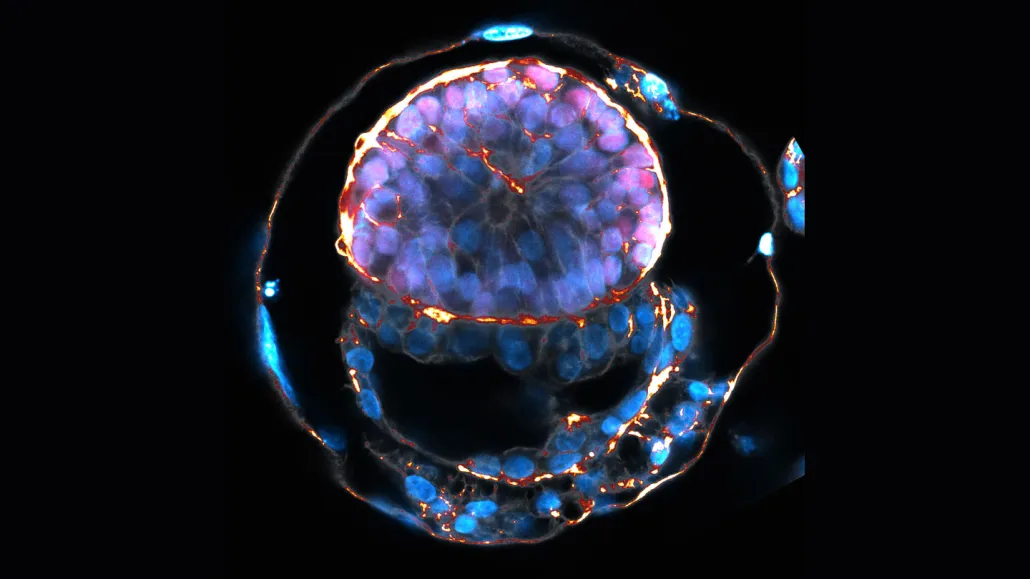
A 6-day-old embryo model from Jacob Hanna’s lab in Israel has structures akin to ones found in human embryos 12 days after fertilization. Those structures include a ball of cells (top, purple) that resemble the layer that forms the body, and a hollow circle of cells (bottom, blue) mimicking the yolk sac, all surrounded by a layer of cells similar to a placenta-forming layer in human embryos.
Jacob Hanna
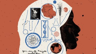
Some newly reported clumps of cells growing in lab dishes have been hailed as the closest things to human embryos that scientists have ever made in the lab.
These entities are human embryo models — masses of cells created from stem cells that mimic some properties of certain stages of embryo development. The achievement gives researchers a chance to look at human development beyond the first week or so, when an embryo must implant in the uterus to develop further. That post-implantation stage hadn’t been re-created in lab dishes — until now.
Six studies reported in June and July describe the embryo models, which have generated excitement and concern in equal measure.
For researchers working on these embryo models, the faux embryos are new tools to gain insight into the “black box” of human development, after embryos implant in the uterus. They are useful because donated human embryos are in short supply, and there are limits on the types of experiments researchers can perform on them.
About 60 percent of pregnancies fail just before, during or soon after implantation, developmental and stem cell biologist Magdalena Żernicka-Goetz of the University of Cambridge and Caltech said June 27 during a news briefing discussing an embryo model made in her lab. Insights gleaned from the embryo models may give new understanding of why many pregnancies fail to take hold and lead to better fertility treatments, Żernicka-Goetz said.
But others worry that the models — along with eggs and sperm made from stem cells — raise the specter of researchers using the mimics to create babies. Scientists developing the models say reproduction is not their aim or intention, and that implantation in a uterus is impossible with these embryo models.
Still, the research raises issues of how to — and whether to — regulate what scientists can do with embryo-like entities made from stem cells. Questions surround whether embryo models could or should be grown past the equivalent of 14 days of normal human development after fertilization. And critics warn that overstating what the models are or can do could risks damaging trust in science.
Science News talked to scientists and ethicists to learn more about these human embryo models.
What are human embryo models?
Before answering that question, Amander Clark, president of the International Society for Stem Cell Research, says we first need to understand that a human embryo is the product of fertilization of an egg and sperm.
Embryo models, on the other hand, self-assemble from pluripotent stem cells — ones that have the power to make nearly any type of cell in the body. “Therefore, embryo models do not meet the clinical, medical or scientific definition of an embryo because they do not originate from the product of fertilization by two gametes,” says Clark, a stem cell scientist, developmental biologist and geneticist at UCLA.
For years, scientists have studied the first week or so of human development using donated human embryos or embryo models (SN: 1/5/22). From those, researchers learned a great deal about the formation of the ball of cells known as a blastocyst. Blastocysts have an outer layer of cells that will form the placenta and other support systems for the developing embryo, and an inner cluster of cells that will give rise to the body.
But it’s the next few weeks of life when the real action happens, says stem cell biologist and embryologist Jacob Hanna of the Weizmann Institute of Science in Rehovot, Israel. Between day seven and about day 35 after fertilization is when the embryo builds all its organs. “It moves from a ball of cells to a structure that anybody on the street … would tell you, ‘This is an embryo.’ And then the rest of the other eight months are just growth of the embryo,” he says.
Researchers have assembled embryo models that contain some, but not all, of the types of cells necessary for normal development. The newly reported embryo models mimic structures that would be found in an embryo that has implanted in a uterus, even though the mimics have nothing to implant into. The models are simulating a very specific window of embryo development: “the events that occur as an embryo implants, how the embryo self-assembles,” Clark says. “Then essentially they collapse.”
None of the models completely copy a real embryo, she says. For instance, none make a very good trophectoderm, the layer of cells that gives rise to the placenta later in development. That layer is important for an embryo to implant into the uterus, and it also sends signals that help the rest of the embryo develop properly.
How did the latest buzz about embryo models start?
Post-implantation embryo models drew media attention when Żernicka-Goetz presented preliminary findings in the closing moments of a talk she gave June 13 in Boston at a meeting of the International Society for Stem Cell Research. The Guardian newspaper hailed the work as a breakthrough that created synthetic human embryos.
That characterization overstates the achievement, says Alfonso Martinez Arias, a developmental biologist at Pompeu Fabra University in Barcelona. The work is not a breakthrough, but an incremental advance, and is far from re-creating an embryo, he says.
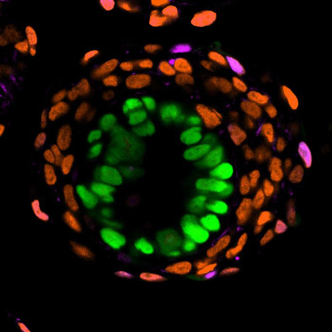
Żernicka-Goetz and colleagues genetically engineered human stem cells to resemble three types of cells necessary for embryo development: cells that mimic the important placenta-generating trophectoderm, cells that resemble ones that will form the yolk sac that feeds the embryo until the placenta takes over, and ones that form the epiblast — the cells inside the embryo that will develop into the body. The resulting balls of cells resemble some aspects of human embryos, the team reported June 27 in Nature.
For instance, the trophectoderm layer forms on the outside of the embryo-like structure as it does in embryos. But it doesn’t make proteins typical for that layer when assembled in a 3-D structure, so it isn’t truly a placental precursor, coauthor Bailey Weatherbee of the University of Cambridge said during the news briefing. But the layer is necessary for the rest of the embryo model to assemble, suggesting it performs some of the functions of the trophectoderm, she said.
How were other embryo models made?
Like Żernicka-Goetz’s team, all the research groups started with human stem cells. Carefully controlling growing conditions and the numbers of certain types of cells added to the mix allowed the stem cells to grow into embryo-like structures. The models differ in the number of cell types they contain and in the features of real embryos they’re able to mimic.
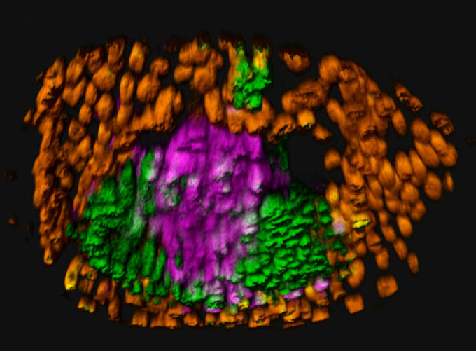
One group of researchers coaxed stem cells to form embryo-like structures with two tissue layers. That work, also described June 27 in Nature, didn’t use genetic manipulation or chemicals to induce the stem cells to form embryo-like structures. Instead, the researchers relied on stem cells’ ability to self-organize, says developmental biologist Berna Sozen of Yale School of Medicine.
The resulting model lacks the trophectoderm. When the researchers realized the tissue was missing, they thought its absence might tell them something about its importance, she says. “In the absence of these tissues you can see what will happen, what doesn’t happen, and then you [get] very strong scientific insights [about] why you need that tissue.”
Another effort by researchers in China and Michigan, described in a not-yet-peer reviewed preprint posted June 16 online at bioRxiv.org, is technically impressive, Martinez Arias says. But it still lacks a placenta precursor layer.
Most recently, a team led by Jun Wu of the UT Southwestern Medical Center in Dallas persuaded stem cells to form embryo-like entities that span the rearrangement event called gastrulation, the researchers report July 20 in Cell. During gastrulation embryos go from hollow spheres of cells to multilayered structures that will give rise to organs and tissues that form the body.
These “peri-gastruloid” models made some tissues resembling those in the early nervous system and may help confirm the origins of cells that will give rise to eggs and sperm. These models also contain a yolk sac, but “our model is not a complete model,” Wu says. “We don’t have the placenta tissue.”
Because the models lack the trophectoderm, the researchers had to add proteins to anchor and support the gastruloids so they could develop properly.
A team of researchers in China reported preliminary results of similar gastruloids with yolk sacs June 28 at bioRxiv.org. That team found that the chemical thalidomide alters formation of the tissue layers and interrupts development.
In the future, researchers might use such models to understand how chemicals from the environment could affect developing embryos, Wu says.
Martinez Arias and others have previously made gastruloids that could model development up to day 19 after fertilization, but those earlier models didn’t have yolk sacs.
Are any of the embryo models major advances?
Of this “gold rush” of embryo models, only two are real advances, Martinez Arias says: Wu’s gastruloids and embryo models made by Hanna’s group.
Hanna’s models described in a June 15 preprint posted at bioRxiv.org, have a reasonable facsimile of both a yolk sac and the placenta precursor, Martinez Arias says, and make structures with “uncanny” resemblance to those of embryos at 14 days of development.
The process to achieve an embryo model so closely resembling the real thing was a long one, Hanna says. His team first worked out how to grow mouse embryos past this developmental stage in lab dishes. Using mouse embryos as their “experimental compass,” the researchers then figured out how to assemble a mouse embryo model from stem cells. From there, the team used the tricks they’d learned to grow human stem cells in conditions that coaxed them to self-assemble into structures approximating post-implantation embryos.
The self-assembly bypassed the blastocyst stage and moved directly to something akin to a post-implantation embryo, his team found. “Maybe the embryo [models] are deficient because we don’t go to through the blastocyst [stage]. I don’t think so. But I can’t exclude it at the moment,” Hanna says.
Can these embryo models result in babies?
No, but that question always comes up.
Scientists are interested in technical and biological advances the new embryo models bring. But for most people, “it’s really the possibility that they could be used for reproduction that that draws the attention,” says Katie Hasson, associate director of the Center for Genetics and Society, a nonprofit social justice organization based in Berkeley, Calif.
It’s not possible to use these embryo models for reproduction, scientists say. “Not only is it illegal to put these late stage [embryo models] inside the uterus, but actually if I wanted to, or anyone wanted to, these structures can never implant,” Hanna says. Implantation happens only when embryos consist of one to 64 cells. These post-implantation models have moved beyond that stage. Biologically, he says, “it will never succeed.”
Wu agrees. “These models are not human embryos at all,” he says. “They are not able to generate any type of life. They are essentially just a cluster of cells.”
There are legal blocks to growing embryos in the lab past a certain stage as well. In the United Kingdom, the law forbids growing embryos past 14 days of development, the point at which the embryo turns on its body-building program. That limit was imposed shortly after in vitro fertilization became possible.
But some of these embryo models already resemble embryos at the 14-day stage of development. Since the models aren’t created through fertilization and could never give rise to a person, some researchers argue that they shouldn’t be subject to the 14-day rule.
In the United States, there is no law banning growing embryos past 14 days, but the 1995 Dickey-Wicker amendment prohibits the U.S. National Institutes of Health from funding research on embryos or embryo-like cells with “organismal potential,” Clark says. Since these models lack critical parts, they have no potential to make an organism — so research with embryo models can be funded.
The international stem cell society recently updated its guidelines, suggesting that if scientists want to culture embryos created by fertilization for longer than 14 days, they need to seek public approval in their jurisdiction. Since the models are not embryos, the society’s guidelines suggest that they can be cultured up until a structure called the primitive streak appears, signaling that the embryo is starting to build the body.
The goal for most scientists in the field is to understand the process by which cells assemble an embryo and then create all the organs and tissues of the body, Martinez Arias says. It’s neither necessary nor desirable, and perhaps not even possible, to build a perfect replica of an embryo. “I certainly do not think that that is something that we want to do, or that we should do or should say,” he says. “And that’s not something that is on the horizon.”
Even so, scientists need to hit the pause button to thoroughly evaluate the embryo-like structures and determine how they can aid in understanding human development, Martinez Arias says. “I hope that now we can all develop these systems in a manner that is useful for research, rather than trying to see how far into space we [can] go, without thinking [about] how we return.”
Conversations with the public about what the embryo models represent scientifically, philosophically and legally, and about how to use them ethically need to start now, Hasson says. “We need to think about them now because it’s not the case, and it shouldn’t be the case, that the technical limits of what scientists can currently do with these models should define the ethical discussion and what the ethical limits should be.”