A new imaging tool could enable researchers to get three-dimensional images of single living cells without resorting to the time-honored procedure of staining their inner structures with chemicals.
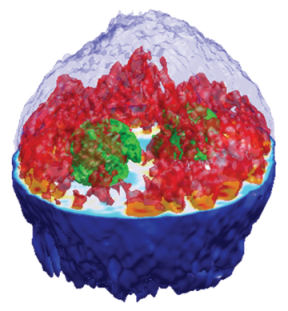
“We can image the cell as it is,” says Wonshik Choi of the Massachusetts Institute of Technology (MIT).