With a new body mapping technique, mouse innards glow with exquisite detail
Creating spongelike holes in mouse tissue helps fluorescent markers penetrate an entire mouse
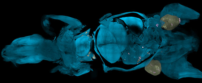
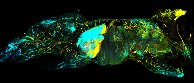
Fluorescent antibodies light up the nervous system of a dead, transparent mouse, lying on its back with its head to the left. Colors show how deep nerve cells are in the animal, from blue (closest to the camera) to pink to yellow (farthest away).
© Ertürk Lab/Helmholtz Munich
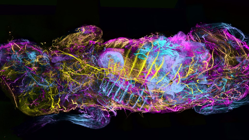
A mouse’s entire nervous system lights up in psychedelic hues. Clumps of immune cells attacking tumors give off a ghostly glow. The vessels that make up the body’s sewer system flare brightly.
These and other images are shining a new light on the inner workings of mice, thanks to a new technique that makes whole body imaging cheaper and faster, researchers report July 10 in Nature Biotechnology.
See-through mice are nothing new (SN: 8/14/14). But existing techniques to image their insides can be expensive, time-consuming or don’t hit the right target. Now, a study shows that chemically removing cholesterol — an essential component of cell membranes — from dead mice creates spongelike holes in tissues without destroying them. That means tailored antibodies can move through the holes to infiltrate every corner of the body and bind to proteins of interest to make entire anatomical features visible under fluorescent light.
The technique, dubbed wildDISCO, gives scientists an extraordinarily thorough peek under mouse skin to create body-wide anatomical atlases. It’s a bit like Google Maps, says Ali Ertürk, a neuroscientist at Helmholtz Munich. But in place of cars driving around to record every street, antibodies act as streetlamps to illuminate scientific landmarks.
Such maps could help researchers train artificial intelligence programs to simulate bodily processes in mice. The algorithms would aim, for example, to simulate how a drug travels through blood vessels, or predict the path of genetically engineered immune cells for cancer therapy, Ertürk says (SN: 2/2/22). Computational simulations of biology may help researchers move away from doing animal experiments, he says.
To make the mice glow, Ertürk and colleagues pumped cholesterol-removing chemicals along with antibodies tagged with fluorescent molecular markers into the dead animals via the heart and blood vessels, a proven tactic that allows liquids to traverse the entire body. Then the team made all the tissues transparent, and the mice were ready for their close-ups.
One of the resulting maps depicts a comprehensive view of the network of nerves that runs beneath the skin and around organs. Another shows off the lymphatic system, an open network of organs and lymphatic vessels that helps fight off pathogens and transport cellular waste for removal.
These blueprints yielded some scientific nuggets.
Images taken of the nervous systems, for instance, showed that mice with no microbes in their bodies had underdeveloped nerve networks in their guts compared with normal mice, suggesting that the gut microbiome plays an important role in helping nerves develop.
And in mice with cancer, the team documented clumps of immune cells inside and near the tumors. The role that these clusters play in controlling cancer is unclear, Ertürk says. But wildDISCO offers scientists a way to investigate.
wildDISCO might also help with studies that focus on animals other than mice, says Yijun Su, an imaging specialist at Howard Hughes Medical Institute’s Janelia Research Campus in Ashburn, Va., who was not involved in the work. Scientists working with fruit flies, frogs or zebra fish might use a similar “recipe,” he says, to take clearer snapshots of their anatomy.