White matter scaffold offers new view of the brain

Neural map may explain why some injuries are worse than others

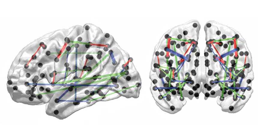
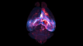
BRAIN TRUST  These white matter tracts (indicated by red, blue and green lines) in the brain are particularly vulnerable to injury, a new study suggests.

A. Irimia and J. Van Horn/Frontiers in Human Neuroscience 2014

Buried amid the complexity of the human brain, a newly described scaffold carries important messages from one place to another. A map of the scaffold, which reveals intricate connections made by bundles of nerve fibers called white matter tracts, could help explain why some brain injuries are particularly devastating.

The scaffold, described February 11 in Frontiers in Human Neuroscience, is distinct from other descriptions of neural connections in the human brain (SN: 2/22/14, p. 22). Instead of focusing on gray matter — swaths of nerve cells themselves — the new network focuses more on their connections. The two approaches are “different ways of getting at the same problem,” says neuroscientist Olaf Sporns of Indiana University Bloomington, who was part of a team that in 2011 described a series of highly linked brain regions called the rich club network. The new connections scaffold is “a very important extension,” he says.

To uncover it, Andrei Irimia and John Van Horn of the University of Southern California scrutinized the brains of 110 healthy men. Structural MRI scans revealed brain anatomy, and another kind of scan called diffusion tensor imaging revealed the shape and size of white matter tracts. After combining the scans to form a computer representation of a brain, Irimia and Van Horn started mathematically snipping connections to see which ones were most important for the scaffold’s shape.

Their approach was like randomly cutting electrical wires in a house, Van Horn says. “You’d find that sometimes you’d cut a wire that doesn’t really change anything other than the outlet in your office,” he says. “Other times you go and cut another wire, and it shuts off the power to your whole house.”

The method identified the white matter pathways that when cut, caused the greatest upheaval to the entire scaffold. Seemingly small injuries to these important fibers may result in outsized devastation, Van Horn and Irimia propose. Conversely, seemingly catastrophic injuries, particularly those in the front part of the brain, might leave a person relatively unaffected because the injury spares important connections.

One such injury resulted when a tamping rod impaled the front part of rail worker Phineas Gage’s brain in 1848 (SN: 2/22/14, p. 32). He did suffer a stark personality change, but in many other regards, his brain continued to function surprisingly well. “If a similar sized injury to Mr. Gage had occurred in another location in his brain,” Van Horn says, “the damage to his network would have been much more severe.”  

It’s not yet clear whether the connection map can help predict the outcomes of brain injuries or diseases. To find out, scientists need more studies on white matter connections in people with injured brains. But this new view of the brain may lead to a deeper understanding of traumatic brain injuries, multiple sclerosis and other disorders in which brain connectivity is disrupted, Van Horn says.


Laura Sanders is the neuroscience writer. She holds a Ph.D. in molecular biology from the University of Southern California.

More Stories from Science News on Neuroscience

From the Nature Index

Paid Content