Zika virus infects cells that make bone, muscle in lab tests

false-color image of cells in minibrain

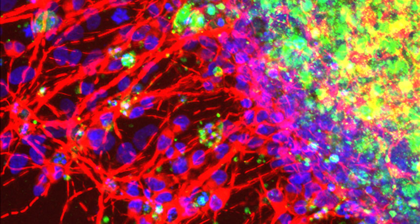
MIXED SIGNALS  Minibrains grown in the lab form nerve cells (red) prematurely and show signs of dying cells (green) when treated with a signaling molecule called LIF. This molecule pours out of embryonic cranial cells after infection with Zika virus, and could harm brain development.

Rachel Greenberg

Zika virus can infiltrate the cells that give rise to bone, cartilage and muscles in the head, researchers report September 29 in Cell Host & Microbe.

In utero infection of these cells, called cranial neural crest cells, could improperly mold babies’ facial features, the authors suggest. The findings — so far observed only in cells and minibrains grown in the lab — offer a possible explanation for the misshapen heads that are the hallmark of microcephaly, a condition that afflicts some babies infected with Zika.

Another hitch: The virus made cranial cells unleash a flood of molecules that can alter brain cell development. So in addition to disfiguring the skull, infected cranial cells might also disrupt the brain, the researchers propose.

Meghan Rosen is a staff writer who reports on the life sciences for Science News. She earned a Ph.D. in biochemistry and molecular biology with an emphasis in biotechnology from the University of California, Davis, and later graduated from the science communication program at UC Santa Cruz.

More Stories from Science News on Health & Medicine

From the Nature Index

Paid Content