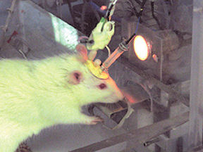
The rat in the plastic box has a drug habit. Every 5 minutes or so, he presses a lever that sends a shot of cocaine through a catheter into his veins. Even more unusual is his “rat hat,” the data-transmitting headgear that monitors the animal’s brain activity without immobilizing the head. The hat positions several insulated wires within the rat’s brain.
For more than 50 years, scientists have been inserting electrodes into tissue samples and animals’ bodies to eavesdrop on electrical activity. But the latest-generation electrodes go further. They detect the ebbs and flows of chemicals at the surfaces of cells.
Researchers first developed electrodes that could measure chemical compounds in the early 1980s, but it took a couple of decades for scientists to figure out the best ways to fabricate and use them, says R. Mark Wightman, an analytical chemist at the University of North Carolina in Chapel Hill.
“Once that was done, people started in on applications,” says Wightman, who is using microelectrodes in rats to study how cocaine influences the brain circuitry that rewards behaviors.
An electrical property of these microelectrodes “enables chemical events occurring on the sub-microsecond time scale to be monitored,” Wightman noted in the March 17 Science.
Moreover, the tools’ small size makes a large difference in their capabilities. The microelectrodes, also called ultra-microelectrodes, are typically 50 to 100 micrometers (µm) long. The wire’s uninsulated tip, which does the sensing, is 10 µm or less in diameter.
With tips in the same size range as cells, microelectrodes are reaching into biological realms that can’t be accessed by their bigger cousins. “If you want to look at chemistry next to a single cell or some other very small space, you would want a microelectrode,” says Wightman.
At the pump
Drugs intended to kill microbes and tumors are often foiled by busy cellular pumps. One research team is using microelectrodes to investigate how cells expel those drugs.
A microelectrode senses what’s going on at the cell’s surface by exchanging electrons with chemicals in solution surrounding the cell. By carefully moving a microelectrode across the surfaces of one or many cells, researchers can map a compound’s concentration.
Allen J. Bard of the University of Texas at Austin applies this technique to cells as they rid themselves of toxic substances. He and his colleagues have used the information from concentration maps in a mathematical model that calculates how quickly a cell pumps out material across its entire surface.
The researchers scanned lab-cultured liver cells as they encountered menadione, an analog of a drug used against cancer. Menadione changes to a toxic form inside the cell. But the cell can tag menadione with another chemical group and pump out the resulting compound, called thiodione, through channels in the cell membrane.
“What we find is that the cell is very efficient at getting rid of the menadione,” says Bard. His group determined that each cell exports 6 million molecules of thiodione per second. That’s almost as fast as the rate at which menadione gets in, he says.
Bard’s group is beginning to study whether cancer cells discharge chemotherapy drugs with a similar speed.
In the long term, Bard’s goal is to shut down the export processes that cancer cells and infectious microbes use to resist drugs intended to kill them. If cells are “using that kind of pump mechanism, and if you could shut that off with another drug, you could get rid of that resistance,” Bard says.
Eyeing cellular exits
Detailed monitoring of cell surfaces can also provide insight into the release of chemical messengers, such as hormones, that carry signals to other cells. Scientists have developed a tool to examine the molecular anatomy of the opening where the cell discharges signal molecules.
During the secretion process, a sac, or vesicle, containing a cargo of molecules inside a cell moves to the cell’s membrane and fuses with it. The cargo then leaves the cell through a channel called a fusion pore that opens between the membranes the vesicle and the cell.
Manfred Lindau, a biophysicist at Cornell University, and his colleagues are working to identify the proteins that form the fusion pore. Toward that goal, they’ve developed a microelectrode array to locate a single active channel. Each microelectrode reports hormone concentration in its vicinity.
The researchers patterned four platinum electrodes onto a glass coverslip, “like a printed circuit board,” Lindau says. In the space at the middle of the four electrodes, they placed a cell that in the adrenal gland releases the stress hormones adrenaline and noradrenaline. The cells, called chromaffin cells, have large vesicles, 200 nanometers (nm) across.
“When you use several electrodes, you can sort of triangulate the position where that release occurs,” says Lindau. His team applied a computer analysis to locate the site secreting adrenaline or noradrenaline.
To confirm that the strategy works, the researchers filled chromaffin-cell vesicles with a fluorescent dye that would flash green when the vesicle spilled its contents. They found that the location indicated by the microelectrode array was within 500 nm of the location shown by the fluorescence. Lindau’s group described the arrays in the Sept. 27, 2005 Proceedings of the National Academy of Sciences.
To learn more about which proteins make up the fusion pore, Lindau and his colleagues plan to attach fluorescent tags to certain proteins. As before, Lindau’s group will place a chromaffin cell in the microelectrode array and locate the secretion event. Then, the researchers will focus on the fluorescence coming from an area as a secretion event occurs there.
Lindau’s team intends to tag three proteins, two found in the cell membrane and one found in the vesicle membrane. The tags’ fluorescence will change only if the proteins change their shape. If this happens at the site of a secretion event in progress, it’s an indication that the proteins are forming a fusion pore.
“We’ll look at fluorescent signals and see how they change related to this fusion-pore opening,” says Lindau, “to give us an idea if there are conformational changes happening.”
Dopamine highs
Installing microelectrodes just outside neurons can illuminate the ways in which specific signal molecules influence behavior. With a microelectrode, “you’re watching in on a conversation of neurons with one another” as they pass information through the brain, Wightman says.
Wightman’s group uses carbon-fiber microelectrodes to detect one chemical message, dopamine. Brain neurons release this neurotransmitter to reward eating, drinking, and some other behaviors critical to survival. Certain addictive drugs also stimulate the release of dopamine.
Past studies have used microelectrodes to detect neurotransmitters in anesthetized animals. But Wightman, psychologist Regina M. Carelli, and their colleagues at the University of North Carolina wanted to learn how dopamine concentrations change in the brains of cocaine-addicted rats as they move around.
So, the university’s machine shop designed the high-tech rat hats that hold three microelectrodes in place in an animal’s brain. To avoid impeding the animals’ normal activities, the researchers ran wires from the hats through a swiveling disk in the ceiling of the plastic box and then to a computer and other equipment. Similarly, the tubing that provides the cocaine permits the rat to wander within the cage.
Two of the microelectrodes placed in the rat brains bookend the groups of nerve cells that constitute the reward pathway. The researchers used one microelectrode to detect dopamine and to measure its concentration fluctuations in the nucleus accumbens, located toward the front of the brain. To stimulate release of dopamine, the researchers used an electrode placed in the ventral tegmental area, which is in the central brain. The third microelectrode was a reference electrode in the cortex on the other side of the brain.
To learn how dopamine concentrations change as the rats inject cocaine, the team fitted six cocaine-addicted rats with the hats. In 2003, they found that about 10 seconds before the rats gave themselves doses of cocaine, dopamine concentrations in the rats’ brains rose for less than a second.
“You can think of it as being an anticipatory response that cocaine is coming,” says Wightman.
In another experiment, the researchers investigated whether they could spur the rats to seek cocaine. They induced an increase in dopamine concentration with the stimulating electrode. Immediately after that, the rats demonstrated the behavioral routines, such as approaching and backing off from the lever, that typically occur before they inject cocaine.
In further work, the researchers detected a dopamine increase about 2 seconds after an animal pressed the cocaine lever. Like the anticipatory burst, this postresponse dopamine increase lasted less than a second.
The researchers sometimes added light and sound cues as a rat received cocaine. The postresponse spike then occurred even when the researchers triggered the flashes and rings without a rat having pressed the lever. In the May 19, 2005 Neuron, Wightman and his colleagues further demonstrated that when rats repeatedly press the lever for cocaine delivery but the researchers block the drug, the dopamine burst that occurs with the cue gradually declines. This suggests that these dopamine spikes teach the rats to associate the cues and the drug, the researchers say.
These transient dopamine changes “would have been absolutely impossible to detect with other techniques,” notes Wightman. “These things all happen right at the lever press, in less than a second—there’s no other way to figure it out.”
He adds that future investigations with this technology should also provide insight into the role of dopamine in behaviors such as learning and memory.
Smaller still
Researchers now plan to move their chemical and electrical measurements from the surface to the inside of cells. To succeed, scientists will have to shrink the sensors further.
Michael V. Mirkin at the City University of New York in Flushing has accepted that challenge. At recent scientific meetings, he has described initial work with a nanometer-size electrode. Its diameter is about one-thousandth that of the smallest microelectrodes now in use.
Scientists have been concerned that putting an electrode inside a cell could cause damage, notes Mirkin, but in his experiments so far, the cell is “essentially not disrupted.” The next step will be to see how well a nanoelectrode provides information from its location inside the cell.
Mirkin says that information collected from experiments using this technique might, for example, have relevance to cancer and other diseases. If it’s possible to use nanoelectrodes in this way, it would be “wonderful,” says Mirkin. But, he cautions, this application “still has to be proven that it works well.”