Defining normal in the brain
Scans set standard for how connectivity evolves during maturation
Researchers have created a growth curve for the brain, similar to the height and weight charts pediatricians use to monitor their patients’ development. Scientists came up with the new developmental milestones by aggregating the results of brain scans that reveal active connections throughout the organ.
Published September 10 in Science, the study reveals how a typical brain’s connections evolve with age, information that could help doctors detect a variety of disorders — many of which are marked by disordered neural connections — earlier.
“It’s really remarkable how much information can be gleaned with just a five-minute resting-state scan,” says neuroscientist Olaf Sporns of Indiana University in Bloomington. “The techniques they’ve developed here may be very powerful in making predictions for individual patients.”
Researchers led by Nico Dosenbach and Bradley Schlaggar, both of the Washington University School of Medicine in St. Louis, constructed the brain maturity curve using data from 238 volunteers ages 7 to 30. Each subject spent about five minutes quietly resting while an MRI, or magnetic resonance imaging, machine recorded patterns of blood movement in over a hundred different brain regions. Because no tasks are required of the patient, the quick scan can be used on infants or patients who are unable to respond to directions.
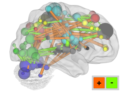
After the data were collected, the researchers fed the brain activity information for each person to a computer, which assessed hundreds of features simultaneously and spit out a score reflecting the “brain age” of the subject. This score was based on how activity in each region of the brain correlated with the activity in all the other regions. In this way, the researchers described the properties of brain connectivity for each of the 238 subjects, and constructed a curve showing how this score goes up over the years.
“If you take your kid to a pediatrician, they plot their head circumference or their height or their weight on a curve,” Schlaggar says. “What we’ve done here is made a growth curve, but in this case, each point reflects the aggregate of 200 different dimensions. We can say, ‘This is the scan of somebody who has a maturity index of 0.8,’ and have a pretty good idea that they are in a normal distribution for their chronological age.”
Overall, the strength of connections doesn’t necessarily increase as the brain matures, but the score does. One reason for the change in score is that links between brain regions that are physically close to each other get weaker with age, while specific long-range connections tend to get stronger.
Connections involving two particular regions — the right anterior prefrontal cortex and the precuneus — were the best predictors for overall brain maturity, the team found. Neither region was a complete surprise: The prefrontal cortex is important for sophisticated cognitive control, including regulating behavior, adapting to new tasks and planning for the future. The precuneus is known to be a major hub for relationships between separate brain regions.
The new study is “a wonderful reminder that the brain really is a network,” Sporns says. “The brain is not just a big ball of connections where nobody knows what’s going on in there. There is structure, which can be discerned with these network tools.”
The researchers don’t envision brain scans becoming a routine part of a pediatric checkup. “That will only happen when the cost of clinical scanning becomes similar to what it takes to measure someone’s weight — and that won’t happen anytime soon,” Schlaggar says.
Rather the test, by uncovering when and how brain maturity deviates from the norm, could be used to help diagnose and track neurological disorders such as Tourette’s syndrome, autism or schizophrenia. Relating the course of disease progression to specific connection deficits could also lead doctors to develop more personalized and effective treatments, Schlaggar says. “Based on this approach, you might be able to predict not just who will have a hard time, but also who will respond to which therapy.”