X-rays reveal what ancient animal mummies keep under wraps
A new method of 3-D scanning mummified animals reveals species IDs and causes of death

Three-dimensional scans of three animal mummies from Swansea University’s Museum of Egyptian Antiquities (a snake (left), bird (top right) and cat (bottom right)) have revealed the mummies’ identities and other secrets. The mummies have not been radio carbon dated, but are likely at least 2,000 years old.
Swansea University
Egyptian animal mummies can look like little more than bundles of cloth. Now high-tech X-rays have unveiled the mysterious life histories of three of these mummies — a cat, a bird and a snake.
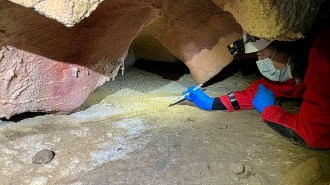
While 2-D X-rays of each specimen existed, little information existed beyond generic animal labels. So Richard Johnston, an engineer at Swansea University in Wales, and his colleagues used a microCT scanner to see what lies beneath the wraps of animal mummies at the university’s Museum of Egyptian Antiquities.
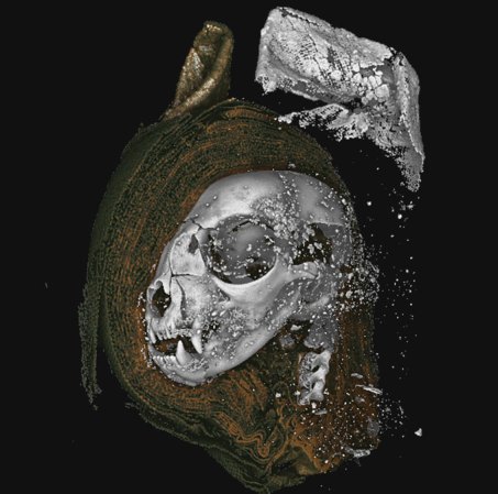
The bone scans of three of those specimens provided such detail that researchers could identify the cat as a domestic kitten (Felis catus), the bird as a Eurasian kestrel (Falco tinnunculus) and the snake as an Egyptian cobra (Naja haje), the team reports August 20 in Scientific Reports. Cause of death was clear in two of the cases: The kitten was strangled, and the snake had its neck broken. The snake also suffered from kidney damage, possibly a result of water deprivation near the end of its life. Like many of Egypt’s mummified animals, these three may have served as offerings to Egyptian gods (SN: 1/6/14).
Focusing on sections instead of just scanning the whole mummy at once allowed the team to get increased detail and create models of the mummified remains that could be 3-D printed and investigated through virtual reality. “With VR, I can effectively make the cat skull as big as my house and wander around it,” Johnston says. That’s how the team found the kitten’s unerupted molars, a clue that the animal was under five months old.
That novel approach to microCT scanning mummies definitely has potential, says Lidija McKnight, an archaeologist at the University of Manchester in England who was not affiliated with the study. “These advanced techniques are extremely powerful tools to improve our understanding of this ancient practice.”
Scans hint at Egyptian ritual in the snake, which had rock structures in its open mouth, possibly the mineral natron used by ancient Egyptians to slow decomposition. Ancient embalmers often opened the mouths and eyes of mummies so the dead could see and communicate with the living, but previously this kind of procedure had primarily been seen in human mummies. Snakes, it appears, may have also whispered beyond the grave, serving as a messenger between the gods and a worshipper.
Peering inside animal mummies with a microCT scanner section by section allowed researchers to create intricate 3-D models with a level of detail that might have been lost through other imaging methods.