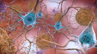
Blood vessels (sans blood) shape organs
It’s obvious that organs maturing in a developing embryo need new blood vessels that will supply oxygen and other vital molecules.
These fledgling blood vessels may do more, however.
Even before blood begins to flow, the vessels provide signals that help give birth to organs, according to two studies of the development of the liver and pancreas. Beyond shedding light on the earliest events in organ formation, or organogenesis, the new results may enable scientists to create artificial organs for transplants or drug-safety testing.
“These are novel and very important findings,” says developmental biologist Deborah Gumucio of the University of Michigan in Ann Arbor. “We have in the past thought of the contribution of the vasculature in terms of delivery of nutrients and oxygen. These studies indicate that before the vasculature is even fully formed, it plays an important role in organogenesis.”
The two studies, which appear in an upcoming Science, reveal that cells lining blood vessels play a key role in the initial growth of the liver in mice and the pancreas in frogs. Development of other organs that require blood vessels for their full function, such as the lungs and kidneys, may also need signals from such cells, suggests Douglas Melton of the Howard Hughes Medical Institute at Harvard University, an author of one of the studies.
“It strengthens the discovery to have two independent groups come up with such similar findings,” says Kenneth S. Zaret of the Fox Chase Cancer Center in Philadelphia, an author on the other study. “This is a cell interaction that may be critical in many more places.”
Zaret’s group investigated liver development in mouse embryos. The team initially noticed that vessel-lining cells called endothelial cells were in close proximity to embryonic tissue that already had begun to express genes known to be active only in the liver. In normal development, this tissue forms so-called liver buds that eventually coalesce into the mature organ.
The researchers then studied mouse embryos that lack endothelial cells because of a genetic mutation. Such embryos never live to birth but do survive to the age at which livers take shape in normal mice. In the mutant embryos, however, there was “a very striking failure of liver-bud emergence,” notes Zaret.
His team then showed that embryonic liver tissue growing in lab dishes stops maturing unless endothelial cells are nearby. The result indicates that the blood vessels provide some developmental signal, either a secreted chemical or a direct cell-to-cell interaction.
“We definitively show the role of endothelial cells at this early stage is intrinsic to the cells themselves and not to their role in creating functional blood vessels,” says Zaret.
Melton and his colleagues have made similar discoveries about the frog pancreas, which secretes hormones such as insulin. They first noticed early blood vessels–without blood–at embryonic sites that would soon develop into pancreatic tissue. Within the immature pancreas, vessels also marked sites where insulin-secreting cells would later develop.
When they placed endothelial cells next to the embryonic tissue from which the pancreas arises, the tissue turned on a pancreas-specific gene called Pdx1 and the gene for insulin. Blood vessels and many organs “develop hand in hand,” concludes Melton. “That guarantees that when you have the final organ, the blood vessels are already there.”
Both his group and Zaret’s are searching for the organ-forming signals that come from the vessels. Meanwhile, Melton is trying to convert embryonic stem cells–laboratory-grown cells with controversial medical promise (SN: 8/18/01, p. 105)–into pancreatic tissue by mixing them with endothelial cells.