Bone marrow in the skull could be used to monitor Alzheimer’s, MS and more
Immune cells in the skull may sense and influence brain inflammation

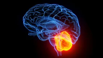
Three-dimensional imaging shows that it’s not just veins (large pink structure at left) that run from the skull and into outer protective layer of the human brain (bottom). Small tunnels (small, blue vertical lines, lower right) also connect this layer to the skull.
Z.I. Kolabas et al/Cell 2023
Cells hidden in the skull may point to a way to detect, diagnose and treat inflamed brains.
A detailed look at the skull reveals that bone marrow cells there change and are recruited to the brain after injury, possibly traveling through tiny channels connecting the skull and the outer protective layer of the brain. Paired with the discovery that inflammation in the skull is disease-specific, these new findings collectively suggest the skull’s marrow could serve as a target to track and potentially treat neurological disorders involving brain inflammation, researchers report August 9 in Cell.
Immune cells that infiltrate the central nervous system during many diseases and neuronal injury can wreak havoc by flooding the brain with damaging molecules. This influx of immune cells causes inflammation in the brain and spinal cord and can contribute to diseases like multiple sclerosis (SN: 11/26/19). Detecting and dampening this reaction has been an extensive field of research.
With this new work, the skull, “something that has been considered as just protective, suddenly becomes a very active site of interaction with the brain, not only responding to brain diseases, but also changing itself in response to brain diseases,” says Gerd Meyer zu Hörste, a neurologist at University of Münster in Germany who was not involved in the study.
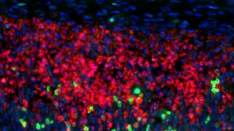
Ali Ertürk of the Helmholtz Center in Munich and colleagues discovered this potential role for the skull while probing the idea that the cells in skull marrow might behave differently from those in other bones. Ertürk’s team compared the genetic activity of cells in mice skull marrow, and the proteins those cells made, with those in the rodent’s humerus, femur and four other bones, along with the meninges, the protective membranes between the skull and the brain.
Cells in the skull, particularly a type of white blood cell called neutrophils, differed from those in other bones but were similar to those in the meninges, the team found. And brain injury widened those differences. In mice with stroke symptoms, the activity in skull cells of genes related to cell migration and inflammation was higher than in other bone cells, suggesting that the cells respond to whatever is happening in the brain.
In another experiment, Ertürk and colleagues compared the profile of proteins in human skull cells with those in the pelvis or the vertebra. Skull marrow cells presented brain proteins while the other bones did not. “This tells us that the communication is two-way,” Ertürk says.
Those discoveries seemed important, but only if the skull and brain had a way to send cells back and forth. A few years ago, researchers observed connecting “tunnels” between the skull and the meninges in mice and humans (SN: 8/31/18). More recently, another team showed that immune cells from the skull were actually getting into the brains of mice, an observation made again in the new study.
To get a better look at the human tunnels, the team used a technique that makes tissue transparent, along with 3-D imaging, to inspect the bone and tissue of people who had died. That revealed a clear path to connect the skull and meninges, something “never been imaged in that much detail in humans before,” says Meyer zu Hörste.
The study “shows the connections very beautifully,” he says, “but how functionally relevant they are still needs to be studied.”
The team did track the location and levels of neuroinflammation in the skulls of people who have Alzheimer’s disease, stroke or multiple sclerosis using PET imaging. The pattern of skull inflammation was different with each disease. For example, in people with MS, there was a huge inflammation signal at the base of the skull, which was not present in people with stroke or Alzheimer’s.
“The inflammatory signature was moving depending on the nature of the neurological disorder,” says Ilgin Kolabas, a neuroscientist in Ertürk’s lab. The signal even increased in specific regions in the skulls of people with Alzheimer’s, a possible sign of the progression of the disease.
Because these signals might be mirroring what is happening in the brain, they could serve as a proxy for neuroinflammation, Kolabas says. The team anticipates these findings could help with both diagnosis and treatment of stroke, Alzheimer’s and other neurological disorders using less invasive methods than spinal taps or other alternatives.
But the technology is still not sufficiently advanced for that yet; future studies need to address whether skull inflammation can actually be measured and targeted to attack brain diseases, Meyer zu Hörste says. “There is a lot of work left to be done.”