New View: Method looks inside embryo fossils

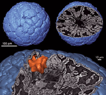
Using an X-ray–scanning technique, scientists have taken a high-resolution peek inside fossilized embryos of some of the earliest multicellular organisms. The procedure offers paleontologists a nondestructive way to see what’s preserved inside ancient rarities smaller than a pinhead and provides fresh insights into the evolution of life on Earth, the scientists say.

INSIDE INFORMATION. The surface-only view (upper left) of a fossil embryo of Markuelia, an ancient relative of marine worms, shows none of the detail visible in a tomographic X-ray image (upper right). The slice at bottom depicts embryonic tissue (white) and a mineral clump (orange). Donoghue, et al.

Bones and shells fossilize more readily than an organism’s soft parts do, but even delicate tissues such as the multicellular embryos in eggs laid by marine organisms can be preserved under the right environmental conditions (SN: 1/28/06, p. 56: Available to subscribers at First Steps).

Previously, scientists had either looked at the surface of a fossil embryo that had been extracted from rock or sliced through the specimen to get a cross-sectional view, says Philip C.J. Donoghue, a geologist at the University of Bristol in England. The first technique provides only limited information, and the latter one is time-consuming and destroys the specimen.

“How to analyze and interpret such fossils is a huge controversy,” he notes.

Now, Donoghue and his colleagues have used a two-story-tall synchrotron to shoot high-energy X rays into ancient embryos of marine organisms no more than half a millimeter across. The team fired the X rays through each specimen at about 1,000 different angles.

A computer then assembled the X-ray images for each specimen into a single, three-dimensional model that depicts features as small as 1 micrometer across. The researchers describe their technique and findings in the Aug. 10 Nature.

When the team scanned the 500-µm-diameter embryo of Markuelia, presumed to be a relative of modern marine worms, the scientists could clearly distinguish the difference between fossilized tissues such as cell membranes and mineral clumps that had infiltrated cavities in the embryo. The image revealed an extra pair of appendages on the organism that wasn’t visible on the embryo’s surface and might have been missed if the specimen had been sliced at the wrong angle, says Donoghue.

The researchers also scanned an embryo of Pseudooides prima, an organism that paleontologists haven’t classified. That image revealed that an enigmatic groove on the organism’s surface extended into the embryo, indicating that the cleft was a biological feature and not a surface defect caused by preservation, says Donoghue.

The odd location of the groove hints that the organism had a previously unknown type of embryonic development, say the researchers. If that were confirmed, scientists could better place P. prima near the base of life’s family tree.

The detail that can be seen in the fossils with the new method is “quite astonishing,” says Derek E.G. Briggs, a paleontologist at Yale University. The technique will prove a boon to interpreting embryonic structures previously hidden, he predicts.

The new findings “add a whole new dimension to understanding the early diversity of fossils,” says James W. Valentine, a paleontologist at the University of California, Berkeley.

More Stories from Science News on Paleontology